
Learn everything you need to know about Dorsal Intercalated Segment Instability (DISI), a wrist condition caused by scapholunate ligament injury leading to pain, weakness, and eventual arthritis if untreated. Our clinic offers expert assessment, dynamic imaging, and personalized rehab to restore wrist stability and prevent progression to SLAC wrist.
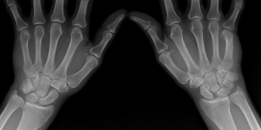
Dorsal Intercalated Segment Instability (DISI) is a specific type of static carpal malalignment where the lunate tilts dorsally (backward) and the scaphoid flexes (forward), leading to a collapsed wrist posture.
Causes of DISI
DISI primarily occurs due to ligament damage, specifically the failure of the scapholunate ligament complex and its secondary stabilizers. More detailed causes include:
Symptoms of DISI
Patients experiencing DISI commonly report a range of symptoms that reflect the instability and altered mechanics of the wrist:
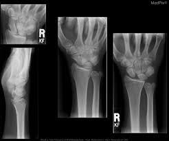
Dorsal Intercalated Segment Instability (DISI) is a static carpal malalignment where the lunate tilts dorsally (backward) and the scaphoid flexes (forward), resulting in a collapsed wrist posture. This specific deformity involves a complex interplay of ligamentous damage and resulting biomechanical failure.
This leads to the radioscaphoid joint becoming incongruent, with the contact area shifting dorsoradially towards the dorsoradial rim of the distal radius. Despite this, congruity is relatively retained in the radiolunate, lunocapitate, and scaphotrapeziotrapezoid (STT) joints. The distal carpal row, including the capitate, trapezoid, and trapezium, also translates dorsally, mimicking the scaphoid's movement due to their intrinsic ligamentous connections. Over time, this abnormal translation and increased contact pressure on the dorsoradial rim of the radius can lead to degenerative arthritis, typically beginning in the radioscaphoid joint.
Dorsal Intercalated Segment Instability is a static carpal malalignment that, if left unaddressed, frequently progresses to degenerative arthritis, commonly known as scapholunate advanced collapse (SLAC) wrist. This progression occurs due to abnormal loading and movement of the wrist, where the dorsoradial translation of the scaphoid can increase contact pressure on the dorsoradial rim of the radius, leading to initial degenerative changes in the radioscaphoid joint. Over time, this degenerative process can also affect the lunocapitate joint, while the radiolunate articulation tends to be preserved. Abnormal lunate and scaphoid positioning in DISI are contributing factors to the development of SLAC wrist.
DISI treatment aims to restore normal carpal alignment and function. These corrective goals include:
Recover faster, move better, and feel stronger with expert physiotherapy. Our team is here to guide you every step of the way.


The fundamental goal in managing DISI is to address the instability and malalignment. This initial phase would conceptually focus on reducing acute symptoms and preventing further progression of the deformity.
Improving Dynamic Stability: While DISI is primarily a static instability visible on radiographs, there are also dynamic scapholunate injury patterns that may not be apparent on standard static imaging. Advanced imaging techniques, such as four-dimensional CT (4DCT), are used to analyze the carpus during various wrist movements like radioulnar deviation, providing dynamic and quantitative analysis of lunate displacement and identifying instability during motion. Correcting the underlying ligamentous and osseous issues aims to restore stable and physiological carpal kinematics during dynamic activities. The goal is to regain functional stability throughout the wrist's range of motion.
Q: Is DISI the same as scapholunate dissociation?
A: Dorsal Intercalated Segment Instability (DISI) is not the same as scapholunate dissociation (SLD), but rather a consequence or deformity that develops from SLD. SLD is the most frequent post-traumatic carpal instability dissociative (CID) type, involving a complete tear of the scapholunate interosseous ligament (SLIL) and often additional tears of secondary ligament stabilisers. This allows the lunate to dissociate from the scaphoid and tend to extend abnormally. Gradually, the DISI deformity develops, which is characterised by the flexion of the scaphoid and extension of the lunate. In DISI, the lunate is in an abnormally extended position in the sagittal plane. This malalignment results in the scaphoid being palmar-flexed and pronated, and the lunate being extended and supinated.
Q: Can DISI be misdiagnosed on imaging?
A: Yes, Dorsal Intercalated Segment Instability (DISI) can be mimicked or misdiagnosed on various imaging techniques, especially on sagittal Magnetic Resonance (MR) images and standard plain radiographs, even in individuals who are asymptomatic and have otherwise normal findings
Q: Does DISI always need surgery?
A: No, DISI does not always require surgery, especially in earlier stages or if it's considered a dynamic instability. However, surgical intervention is often considered for chronic or static deformities. A critical factor for timely intervention, whether non-surgical or surgical, is to prevent the progression of DISI to scapholunate advanced collapse (SLAC) arthritis.
At our clinic, we use:
Book Your Assessment Today:
Serving Vaughan, Thornhill, Richmond Hill, Markham, and North York. Direct billing available.
Key Takeaways:
Explore the latest articles written by our clinicians
.webp)
.webp)
.webp)


.avif)


