Cyclist undergoing professional bike fitting and biomechanical assessment for Iliotibial Band Syndrome (ITBS) rehabilitation in our clinic

Iliotibial Band Syndrome

Musculoskeletal or neurological condition affecting mobility or function.

What Is Iliotibial Band Syndrome?

The Iliotibial Band Syndrome (ITBS) is recognized as one of the most common overuse syndromes causing knee pain and is often referred to as runner's knee.

Based on the sources, here is a comprehensive overview of what Iliotibial Band Syndrome is, its mechanism, and its characteristic symptoms:

Nature and Etiology

ITBS is fundamentally an overuse disorder of the lateral knee resulting from repetitive movements. It is characterized by irritation and inflammation of the iliotibial band (ITB) [Query], a structure that is a complex thickening of the fascia lata located in the lateral part of the thigh, consisting of longitudinally arranged fascial connective tissue.

The etiology of ITBS is controversial and likely involves multiple factors. Several mechanisms have been proposed:

  1. Friction Theory (Traditional View): ITBS is often presumed to be caused by excessive tension in the ITB. Historically, the condition was thought to result from friction of the band as it rubs repeatedly over the lateral femoral condyle (LFC) (the outside of the knee joint) during alternating knee flexion and extension [8, 36, 80, 84, Query]. This friction leads to inflammation of adjacent soft tissues.
  2. Compression/Strain Theory (Modern View): The friction theory has been challenged because the ITB is firmly attached along the linea aspera of the femur by fibrous bands, which prevents significant lateral movement over the condyle. Current prevailing theory suggests that compression is the primary cause of ITBS. This compressive force acts on the highly innervated fatty tissue and soft tissues located deep beneath the distal ITB attachment.
  3. Strain Rate: A prospective study indicated that a major factor in the development of ITBS is a significantly greater strain rate in the ITB during running, suggesting that strain rate, rather than the magnitude of strain or the duration of impingement, may be a causative factor.

Symptoms and Clinical Presentation

The key symptom of ITBS is sharp or burning pain along the lateral aspect of the knee. This pain is localized specifically near the lateral femoral epicondyle, in the area between Gerdy’s tubercle and the LFC.

The presentation of the pain typically follows a progressive pattern:

  • Onset: Pain is correlated with regular physical activity. It often occurs during physical activity after a reproducible time or distance of running.
  • Progression: Initially, the pain may only be felt after a longer duration or upon completion of the athletic activity. However, as the dysfunction progresses, the pain appears earlier during activity or may even occur at rest.
  • Exacerbation: Pain is often exacerbated by activities that increase ITB tension, such as running downhill, lengthening the stride, and descending stairs.
  • Location: Pain is most acute at approximately 30 degrees of knee flexion. The condition may also be associated with a feeling of snapping.

Epidemiology

ITBS is particularly common in the athletic population:

  • It is the most common cause of lateral knee pain in runners and cyclists.
  • The reported incidence ranges from 1.6% to 12% in runners.
  • ITBS is also seen in athletes participating in sports that involve repetitive movements, including tennis, football, skiing, weight lifting, dancing, volleyball, and aerobics.
  • It occurs slightly more often in women than in men. For example, it has been reported as a cause of knee pain in 62% of female runners and 38% of male runners, as well as 24% of cyclists.
  • The syndrome rarely appears in the population of professionally inactive individuals.

Key Symptoms:

The symptoms and diagnostic considerations you listed align closely with the established clinical presentation of Iliotibial Band Syndrome (ITBS), which is considered the most common cause of lateral knee pain in runners and cyclists.

Here is a detailed breakdown of the key symptoms and the necessary differential diagnoses, supported by the provided sources:

Key Clinical Symptoms of Iliotibial Band Syndrome (ITBS)

1. Pain Characteristics and Location

The definitive key symptom of ITBS is sharp or burning pain located in the lateral compartment of the knee.

  • The pain is specifically experienced in the area of the distal part of the iliotibial band (ITB), often localized between Gerdy's tubercle and the lateral femoral epicondyle (LFC).
  • The most acute pain is felt at approximately 30 degrees of knee flexion. This specific angle is critical because it relates to the mechanism of injury (friction or compression).

2. Timing and Progression of Pain

ITBS is an overuse disorder, and the pain typically follows a progressive pattern correlated with repetitive activity:

  • Pain commonly occurs during physical activity, appearing after a reproducible time or distance of running.
  • Initially, the pain may only be felt after a longer duration or after the completion of the athletic activity.
  • As the dysfunction progresses, the symptoms can appear earlier during activity or may even occur at rest.

3. Exacerbating Activities

The pain associated with ITBS is often worsened by activities that increase the strain or friction/compression on the distal ITB:

  • Symptoms often worsen during activities such as downhill running [Query, 10, 87] or when descending stairs.
  • Running on inclined surfaces, such as downhill, is a factor because the knee works in a restricted range of motion, oscillating around 30 degrees of flexion, which increases the frequency of friction/compression of the ITB.
  • Pain is also exacerbated by lengthening the stride.

4. Physical Signs and Sensations

During a physical assessment, specific signs are often present:

  • Tenderness or tightness along the IT band: Palpation of the distal ITB should reproduce the patient’s typical pain symptoms.
  • A clicking or snapping sensation (or "snapping feeling") may be associated with the localized pain near the distal ITB.
  • During a palpation examination conducted alongside movement of the affected joint, crepitations may also be present.

Important to Rule Out (Differential Diagnosis)

The diagnosis of Iliotibial Band Syndrome (ITBS) is primarily achieved through a thorough clinical history and physical assessment. Due to the common nature of lateral knee pain, making a differential diagnosis is critical to ensure the symptoms are not arising from other pathologies.

The sources support the necessity of ruling out several conditions that mimic ITBS symptoms:

Required Differential Diagnoses

Lateral thigh and knee pain can be referred from various conditions, making the exclusion of alternative diagnoses essential for patients suspected of having ITBS. Conditions that must be initially ruled out include:

  • Patellofemoral Pain Syndrome (PFP): This is one of the alternative sources of knee pain that needs to be excluded. It is relevant to note that Patellofemoral Pain Syndrome has been reported to have a higher prevalence among runners than ITBS.
  • Lateral Meniscus Tear/Damage: Damage to the lateral meniscus is among the knee conditions that should be ruled out first.
  • Referred Pain: Pain in the lateral thigh and knee area can be referred from other anatomical regions, meaning that lumbar radiculopathy (nerve root irritation from the spine) and gluteal tendinopathy must also be excluded.
  • Other Local Knee Conditions: Additional conditions to exclude are lateral synovial fold syndrome and distal femoral damage.

Diagnostic Testing and Imaging

While ITBS is typically diagnosed clinically, specific functional tests aid in the physical assessment:

  1. Noble Compression Test: This is a key functional test for ITBS. To perform this test, the examiner applies pressure to the iliotibial band 2 cm proximally to the lateral femoral epicondyle. The knee is then passively moved from 0 to 60 degrees of flexion. A positive result occurs when the patient experiences typical knee pain at 30 degrees of flexion.
  2. Ober's Test: This test is used to assess the passive elasticity of the iliotibial band complex. A positive test leads to pain, compression, or a clicking/snapping sensation of the iliotibial band.

Diagnostic Imaging is generally not required for initial diagnosis but is reserved for cases that are recurrent or resistant to conservative treatment and needs to be correlated with clinical information.

Anatomy of the Iliotibial Band

The Iliotibial Band (ITB) is a critical anatomical structure of the lateral thigh, often implicated in Iliotibial Band Syndrome (ITBS), which is considered an overuse disorder of the lateral knee.

Here is a detailed anatomical description of the ITB, addressing the points in your query, and incorporating insights from the provided sources:

1. Structure and Attachments

The ITB is fundamentally a complex structure located in the lateral part of the thigh, described as a lateral thickening of the circumferential fascia lata that completely envelops the leg. It is composed of longitudinally arranged fascial connective tissue.

  • Proximal Origin (Hip/Iliac Crest): The ITB originates as a sheet of connective tissue originating at the iliac crest. It is formed by the consolidation of fibers from various structures.
  • Muscular Connection: The band is closely connected to and receives fibers from the Tensor Fasciae Latae (TFL), which inserts directly into the ITB. It also receives input from the fascial components of the Gluteus Maximus (a substantial portion of which inserts directly into the ITB) and the Gluteus Medius.
  • Course along the Femur: The ITB runs down the lateral thigh and is firmly attached along the linea aspera of the femur from the greater trochanter up to and including the lateral femoral condyle (LFC). This attachment occurs via coarse fibrous bands or the lateral intermuscular septum.
  • Distal Insertion (Lateral Tibia/Gerdy’s Tubercle): The ITB terminates at Gerdy’s tubercle of the tibia [36, 83, 84, 96, Query]. It ends via multiple attachments in the lateral compartment of the knee joint, including the lateral femoral epicondyle, Gerdy's tubercle, and fibers that articulate with the patella.

2. Role in Knee Stabilization and Movement

The ITB is recognized as a structure that functions like a tendon, consisting largely of dense, regular connective tissue.

  • Stability: Historically, the ITB was considered a ligament connecting the hip and knee to assist in balance in static positions and during movement. Proximally, the ITB acts as a lateral hip stabilizer resisting hip adduction.
  • Load Bearing: Being largely fascial tissue, it exhibits a significant capacity for adaptation and transferring mechanical loads. It has been shown to be an important co-contributor to elastic energy return during running.

3. Crossing the Lateral Femoral Condyle and ITBS Mechanism

The ITB runs down the lateral thigh and passes over the lateral femoral epicondyle (LFE). This interaction point at the knee is central to the etiology of ITBS, characterized by irritation and inflammation [Query].

  • Friction Theory: ITBS has been assumed to result from inflammation secondary to friction of the band across the LFE during knee flexion and extension. The resultant pain is felt specifically at the lateral femoral condyle (LFC).
  • Peak Irritation Angle: Symptoms are commonly associated with the knee joint oscillating around a specific range of motion. Pain is most acute at 30 degrees of flexion. The impingement zone—the range where the distal fibers are believed to compress and slide over the LFE—is thought to occur between 20 and 30 degrees of knee flexion.
  • Modern View (Compression): The traditional friction theory is challenged by anatomical findings showing that the ITB is firmly attached to the femur along the linea aspera via fibrous bands. This attachment prevents significant lateral movement over the condyle. The currently prevailing theory suggests that compression is the primary cause of ITBS development, where the ITB presses on the underlying, highly innervated fatty tissue deep beneath its distal attachment.

Start Your Journey to 

Better Health Today

Recover faster, move better, and feel stronger with expert physiotherapy. Our team is here to guide you every step of the way.

Why It Matters

Iliotibial Band Syndrome (ITBS) matters significantly because if the condition is left unmanaged or poorly treated, it can transition from an acute overuse injury into a chronic, debilitating disorder with measurable tissue changes and a high risk of recurrence.

Here is a breakdown of why ITBS matters, drawing on the consequences of its development and progression:

1. Risk of Chronicity and Progression

ITBS has a tendency to follow a variable course and can recur at any point during treatment or after the athlete returns to full activity.

  • Treatment Resistance: While ITBS is primarily managed non-operatively, the reported cure rate is modest. Cases that are recurrent or resistant to conservative treatment may require diagnostic imaging for further evaluation. In severe cases, surgery is sometimes required to resolve the problem.
  • Worsening Symptoms: If the condition is allowed to progress, the sharp or burning pain symptoms, which initially occur only after a reproducible time or distance of running, can appear earlier during athletic activity or even at rest.

2. Significant Limitation of Activity

As an overuse syndrome highly prevalent in athletes, ITBS directly interferes with the ability to participate in running and cycling.

  • Functional Impairment: ITBS symptoms, which include pain at the lateral aspect of the knee, can negatively impact not only knee function but also psychological and physical aspects of health-related quality of life.
  • Exacerbating Activities: The dysfunction directly limits activities that involve repetitive flexion/extension of the knee, such as running, cycling, and field sports. Pain is notably aggravated during downhill running or descending stairs.
  • Need for Rest: In the acute phase, the first step to alleviate pain is often to refrain from activities that provoke the ailment to avoid the recurrence of symptoms, leading to enforced rest or activity modification.

3. Pathological Changes (Inflammation and Fibrosis)

Repetitive irritation, stemming from friction or, more commonly, compression of soft tissue beneath the Iliotibial Band (ITB), leads to measurable pathological changes.

  • Chronic Inflammation: Gross pathology in tissue obtained at surgery has revealed features of chronic inflammation in the tissue situated between the distal ITB and the lateral femoral condyle (LFC). Local inflammation in this area may be linked to the compression of the richly innervated loose connective tissue.
  • Fibrosis/Thickening: Chronic ITBS has been shown to cause deformation and thickening in the distal part of the ITB.

4. Increased Recurrence Risk

The cycle of injury, inflammation, and pathological thickening contributes to instability and poor mobility, increasing the risk of the condition returning.

  • Preventing Recurrence: To stop ITBS from recurring, eliminating modifiable risk factors is essential. Modifiable factors include training errors (like suddenly increasing distance or running excessively on inclined surfaces) and intrinsic factors like weakness of the hip abductor muscles.
  • Worsened Mobility and Stiffness: To ensure long-term effectiveness of treatment, the compression of the highly innervated tissues beneath the ITB must be reduced. Addressing biomechanical deficiencies through strengthening exercises (especially for the hip abductors and external rotators) is considered a key goal, as impaired hip musculature is often suggested to be related to ITBS.

Causes and Risk Factors

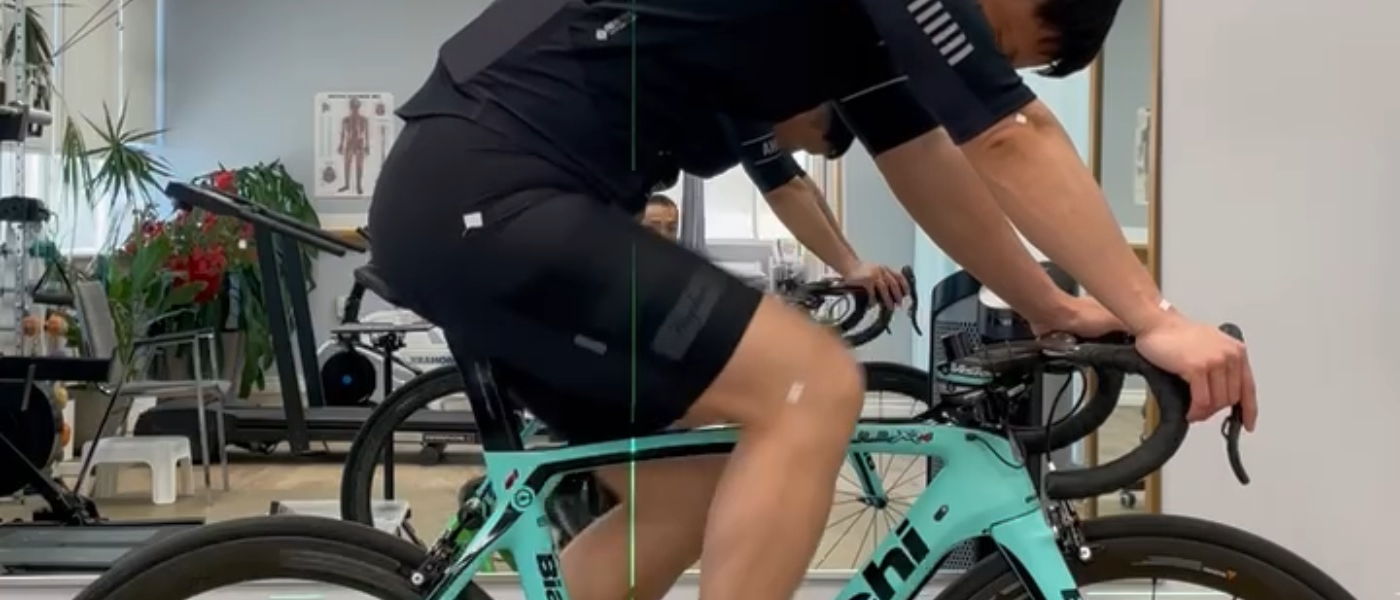
Iliotibial Band Syndrome (ITBS), commonly referred to as runner’s knee, is the leading cause of lateral knee pain in runners and endurance athletes. It is considered a multifactorial overuse injury, primarily resulting from increased tension and strain in the iliotibial band due to repetitive lower limb movements.

Primary Etiological Mechanisms

Historically, the most accepted explanation for ITBS was the friction theory, which suggested that repetitive bending and straightening of the knee caused the ITB to rub over the lateral femoral condyle, leading to irritation and inflammation. However, current understanding favors the compression theory. This model proposes that pain originates from compression of the richly innervated soft tissue and fat pad beneath the distal ITB — particularly during activities involving approximately 30 degrees of knee flexion. Excessive hip adduction and knee varus (bowing) are thought to exacerbate this compressive force.

Additionally, emerging research highlights the role of ITB strain rate — that is, how quickly and frequently the band is loaded during movement. Runners who exhibit higher strain rates in the ITB are more likely to develop ITBS, suggesting a mechanical-biological link in the pathogenesis.

Extrinsic (Modifiable) Risk Factors

Extrinsic risk factors are primarily related to training habits and equipment choices, making them key targets for prevention.

Overuse and training errors are the most significant contributors. Sudden increases in running distance or intensity, especially without proper conditioning, can overload the ITB. Downhill running is another well-documented risk factor, as it places the knee in a constant mid-flexed position (~30°), increasing the frequency and intensity of ITB compression. Runners who accumulate high weekly mileage without sufficient recovery are particularly at risk.

Other extrinsic factors include engaging in interval training, which can overload the lower limbs if not programmed carefully. Poor footwear choices, especially shoes lacking support or worn-out soles, may alter lower limb mechanics and increase ITB stress. Additionally, inefficient running technique — such as overstriding, excessive hip adduction, or increased internal rotation of the knee — can significantly elevate ITB tension.

Intrinsic (Biomechanical and Anatomical) Risk Factors

Intrinsic factors involve a runner’s muscle function, alignment, and anatomical variations, all of which affect how forces are distributed across the hip and knee during movement.

One of the most consistently identified internal risk factors is weakness in the hip abductor muscles, especially the gluteus medius. These muscles are essential for maintaining lateral hip stability and resisting hip adduction. If they are underactive, the ITB compensates by taking on more load, increasing the likelihood of irritation. Weakness in the knee extensors and flexors has also been implicated.

Structural issues such as knee varus (bow-legged posture) or dynamic knee varus during movement can place the ITB under continuous lateral tension. Similarly, excessive femoral adduction, tibial internal rotation, and pronation of the foot can collectively increase ITB strain. One study found that runners with ITBS had significantly greater rearfoot motion (i.e., pronation) compared to healthy controls.

A leg length discrepancy — even a small one — can alter pelvic tilt and gait mechanics, leading to asymmetrical loading of the ITB. Likewise, tightness in the ITB or limited passive hip adduction range of motion can predispose an athlete to ITBS, especially under high loads.

Other intrinsic factors include being under the age of 34, having a history of previous lower limb injuries, and exhibiting an increased knee flexion angle at heel strike during running. Epidemiological data also indicate that females may be slightly more prone to ITBS than males, likely due to anatomical and biomechanical differences (e.g., wider pelvis and increased Q-angle).

This multifactorial profile emphasizes the importance of a comprehensive physiotherapy assessment that not only addresses the symptoms of ITBS but also investigates training habits, movement quality, and underlying biomechanical deficits to reduce recurrence risk.

Why Physiotherapy is Essential

Physiotherapy, which encompasses a comprehensive conservative management approach, is considered essential and is the mainstay of treatment for Iliotibial Band Syndrome (ITBS). Most patients who utilize only conservative management experience complete symptom relief and are able to return to full activity within 4 to 8 weeks.

The treatment plan relies on a progressive, multi-phase approach involving therapeutic exercise, manual therapy, neuromuscular re-education, and modalities.

1. Reduces Pain and Inflammation Through Manual Therapy

The first and immediate goal in the conservative management of ITBS is to alleviate symptoms and eliminate the inflammatory state.

  • Initial Control: Measures used in the acute phase include rest from activity that provokes the ailment, application of ice, and pharmacological intervention such as Non-Steroidal Anti-inflammatory Drugs (NSAIDs).
  • Targeting Irritation: The pain of ITBS is now thought to be primarily related to the compression of the richly innervated fatty tissue beneath the distal ITB. To ensure the long-term effectiveness of treatment, this compression of nociceptive tissues must be reduced.
  • Manual Techniques: Manual therapy is used, though specialized studies are limited. These techniques should address myofascial restrictions, active trigger points, and lumbopelvic dysfunction. Specific soft tissue techniques, such as myofascial release using a foam roller, are incorporated to counteract myofascial adhesions and increase mobility of the tissue complex. The use of an osteopathic manipulative technique called counterstrain has also been shown to improve function and alleviate pain in a case study of ITBS.
  • Modalities: The application of local cryotherapy and other physical therapy modalities like phonophoresis and iontophoresis further supports the reduction of inflammation and symptoms during the acute phase.

2. Improves Glute and Hip Strength to Reduce IT Band Strain

A crucial element of long-term ITBS rehabilitation is addressing the underlying biomechanical deficiencies that cause excessive strain and tension on the ITB. Weakness of the hip abductor muscles is cited as a major intrinsic risk factor for ITBS.

  • Focus on Hip Stabilizers: Individually tailored hip strengthening is a key component of ITBS rehabilitation. Exercises concentrate on improving the strength of the muscles of the lower limb girdle.
  • Reducing Strain: The ITB acts as a lateral hip stabilizer, resisting hip adduction. Impaired hip musculature is suggested to be related to ITBS. Strengthening the hip abductor muscles—particularly the gluteus medius and gluteus maximus—aims to reduce femoral adduction and internal rotation during loading activities, thereby decreasing strain rate and compressive forces on the ITB.
  • Exercise Progression: Strengthening programs, such as the "progressive model 3-phase" scheme, emphasize exercises like "clamshells," short-arc hip abduction, and single-leg hip raises, which focus on improving the endurance of hip girdle muscles.

3. Identifies and Corrects Biomechanical Faults and Re-establishes Proper Movement Patterns

Physiotherapy is essential for identifying and correcting biomechanical factors, as these are often the root cause of the increased strain rate that leads to ITBS.

  • Neuromuscular Re-education: Treatment involves neuromuscular re-education of movement patterns. This aims to restore more desirable movement patterns, ensuring high quality and controlled movements.
  • Controlling Alignment: Exercises should be performed with high quality of movement to control excessive internal rotation and adduction at the hip joint. For example, closed-chain exercises, such as unilateral lunges and half-squats, require the hip abductor muscles to prevent the pelvis from dropping on the contralateral side (Trendelenburg sign).
  • Phased Rehabilitation: The rehabilitation process follows a structured progression:
    • It begins with open chain exercises with low loads to correctly activate the hip girdle muscles.
    • It progresses to closed chain exercises with moderate loads, where endurance gradually increases, and mirror feedback may be used to improve the control and quality of movement.
    • The final phase includes dynamic activities like unilateral deep squats and jumping sequences to confirm tolerance and readiness for return to full activity.

4. Prevents Recurrence Without Need for Injections or Surgery

Conservative management is the preferred path, as it addresses the cause rather than just the acute symptoms, thereby reducing the risk of chronicity and avoiding invasive procedures.

  • Addressing the Root Cause: While local treatment measures (like injections or NSAIDs) may be useful for temporarily easing acute symptoms, addressing the underlying biomechanical basis is more likely to result in long-term benefit and is crucial for preventing recurrence.
  • Preventing Recurrence: Eliminating modifiable risk factors, such as training errors (e.g., sudden increase in mileage or excessive downhill running), is key to preventing ITBS from recurring.
  • Avoiding Invasive Treatment: ITBS is generally treated non-operatively. Surgical intervention is rare and is only required in some cases to resolve the problem. Successful physiotherapy ensures that patients can return to full activity based on tolerance for progressive loading and absence of pain.

Prognosis: Recovery Timeline

The outlook for Iliotibial Band Syndrome (ITBS) is generally positive with consistent, conservative management. However, the recovery course can vary between individuals, and symptoms may recur if the underlying causes aren’t addressed.

General Recovery (Typical Cases)

  • Most cases resolve with conservative treatment (physiotherapy, rest, and activity modification).
  • Full return to activity is typically achieved within 4 to 8 weeks.
  • Key treatment focuses:
    • Reducing inflammation and pain (e.g. rest, ice, NSAIDs, soft tissue work).
    • Improving ITB flexibility through stretching and release techniques.
    • Strengthening hip girdle and core muscles to reduce ITB strain.
    • Correcting training errors and running/cycling biomechanics.

Chronic or Recurrent Cases

  • Chronic ITBS is typically defined as symptoms lasting 3 months or more.
  • Pain may:
    • Occur earlier during activity or persist at rest.
    • Become recurrent if the root biomechanical issues aren't corrected.
  • These cases often involve:
    • Persistent biomechanical dysfunction, such as poor hip control or excessive hip adduction/internal rotation.
    • Underlying weakness in the hip abductors and stabilizers.
    • Poor movement quality during running or functional tasks.
  • Treatment must include:
    • Advanced biomechanical correction and movement retraining.
    • Neuromuscular re-education to improve control and coordination.
    • High-quality strength work with focus on form, alignment, and endurance.
    • Gradual re-introduction to sport or training volume with monitoring.
  • In resistant cases:
    • Imaging (e.g. MRI or ultrasound) may be considered to rule out other conditions.
    • Referral for medical management may be necessary if conservative care fails.

Physiotherapy Treatment Plan

Conservative management is widely supported as the mainstay of treatment for ITBS. The most effective approach follows a phased, multi-modal rehabilitation model progressing through three overlapping phases:

  1. Acute (Pain Relief & Mobility)
  2. Subacute (Flexibility & Muscle Activation)
  3. Strengthening & Return to Sport

This strategy focuses on reducing excessive tension and compression of the richly innervated tissues beneath the distal ITB—the key cause of pain in ITBS. With a well-structured rehab plan, most patients return to full activity within 4 to 8 weeks.

Phase 1: Pain Relief & Mobility (Acute Phase – Weeks 0–2)

The initial phase aims to reduce inflammation and tissue sensitivity by limiting aggravating activities and alleviating mechanical irritation.

  • Relative rest is essential. Temporarily stop activities that provoke symptoms (like running or cycling) to allow inflammation to settle before progressing into active rehab.
  • Ice or cryotherapy can be applied locally during acute flare-ups to reduce pain and inflammation.
  • Manual therapy should address surrounding myofascial restrictions. Techniques like myofascial release or trigger point work are most effective when directed at the TFL, gluteus maximus, and related structures—muscles directly contributing to ITB tension.
  • Gentle stretching of the ITB–TFL complex may help relieve tension, especially in cases with reduced range of motion. Stretching should begin early in the rehab program if tolerated, using positions like lateral trunk flexion with hip adduction (held for ~30 seconds, repeated 3 times).
  • Modalities such as NSAIDs, phonophoresis, or iontophoresis may provide additional pain relief in the acute stage.

Phase 2 & 3: Muscle Activation, Strengthening & Motor Control (Weeks 2–8)

These stages focus on correcting the underlying dysfunctions that caused ITBS, particularly hip weakness, poor movement control, and excessive strain rate through the ITB.

  • The primary goal is to activate and strengthen the hip abductors (especially gluteus medius and maximus) and core stabilizers. Weakness in these muscles is a well-established intrinsic risk factor for ITBS.
  • Early in this phase, begin with low-load open chain exercises to promote motor control. Effective options include clamshells, side-lying hip abduction, and isometric holds with proper alignment.
  • Foam rolling (self-myofascial release) is used to improve soft tissue extensibility, particularly in the ITB–TFL complex. Rolling from the greater trochanter to just above the lateral knee joint line, for 1–5 minutes per session, has shown positive short-term effects on pain and flexibility. Spending at least 2 minutes on the painful area is generally recommended.
  • As pain decreases and strength improves, progress to closed chain and single-leg exercises, such as unilateral lunges, half-squats, step-downs, and single-leg bridges. These help retrain strength and stability through functional movement.
  • Neuromuscular re-education is critical to restore proper movement patterns. Poor control of hip adduction and internal rotation increases ITB strain. Use mirrors or video feedback to reinforce high-quality movement and alignment during all exercises.

Phase 4: Return to Sport & Maintenance (Week 8+)

Progression to this phase depends on the ability to walk for 30 minutes and run for 1 minute pain-free.

  • Begin a gradual return-to-run protocol, starting with alternating intervals of running and walking on flat, predictable terrain (e.g., 1 minute running, 1 minute walking). Run every other day at first, progressing to daily running based on tolerance.
  • Avoid hills or uneven terrain until the athlete is fully pain-free with moderate distances on flat ground.
  • Incorporate gait retraining as needed. Common errors—such as overstriding, hip drop, or excessive knee internal rotation—should be addressed by a trained clinician to reduce re-injury risk.
  • Introduce dynamic loading and plyometric drills: deep single-leg squats, jumping progressions, lateral bounds, and agility ladder work are useful for reconditioning tissue for sport-specific demands.

Prevention & Long-Term Management

Preventing recurrence of ITBS requires ongoing attention to modifiable risk factors, biomechanics, and loading patterns.

  • Manage training loads carefully. Avoid sudden increases in mileage or intensity, and limit frequent downhill running, which increases ITB strain due to repetitive flexion at ~30°.
  • Continue strengthening the hip abductors and core. These muscle groups must be maintained with consistent exercise to provide dynamic control of hip and knee alignment.
  • Incorporate regular flexibility and soft tissue work—foam rolling, stretching, massage, and, when appropriate, dry needling—to address soft tissue tightness that may predispose to ITB tension.
  • Wear appropriate footwear for your foot type and replace shoes before they wear down, especially if you're running high weekly mileage.
  • Periodically review running form and bike fit (for cyclists), as faulty mechanics like excessive foot pronation, overstriding, or poor pedal alignment can contribute to recurring ITB irritation.

By adhering to a phased, evidence-based rehabilitation plan, and addressing the root causes of ITB overuse, most patients can expect a complete and lasting recovery from Iliotibial Band Syndrome. Consistency in strength, mobility, and form maintenance is essential for long-term success.

Ready to Get Back to Running Pain-Free?

Our Specialized Approach to IT Band Syndrome Rehab

We offer:

  • Hands-on release techniques for ITB and hip tightness
  • Targeted gluteal and core strengthening
  • Running and movement analysis to fix biomechanical drivers
  • Personalized rehab programs for fast and lasting recovery

Book Your Assessment Today:

Written by Emilie Yau

Team

Expert Insights

Explore the latest articles written by our clinicians