Golfer swinging club with risk of medial epicondylitis (golfer’s elbow) from repetitive arm motion

Medial Epicondylitis (Golfer’s Elbow)

Pain or dysfunction involving the elbow joint.

Medial Epicondylitis (Golfer's Elbow) Rehab Guide

What is Golfer’s Elbow?

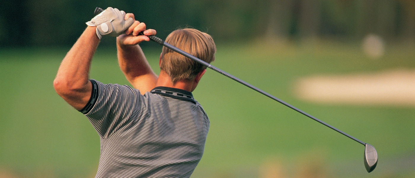
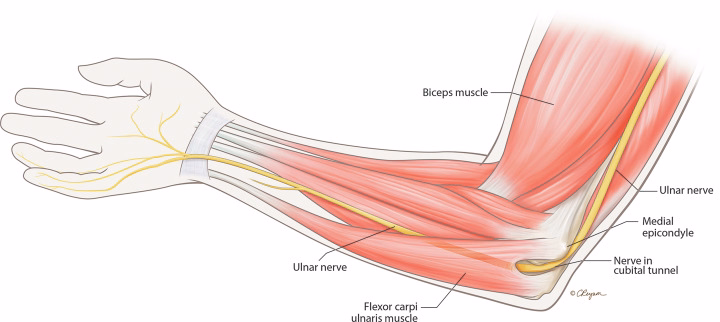
Definition:Medial epicondylitis (ME), commonly known as “golfer’s elbow,” is an overuse syndrome of the elbow. It specifically involves injury to the flexor-pronator group (FPG) of muscles, which is also referred to as the common flexor tendon (CFT). This injury occurs where the FPG attaches to the medial epicondyle of the humerus.

The primary mechanism of injury is repeated eccentric loading of the FPG muscles. This often happens during activities that combine wrist flexion and forearm pronation with a valgus force on the elbow, such as those performed by overhead throwing athletes or manual laborers. Repeated stress leads to microtrauma and degeneration of the musculotendinous units. While the suffix "-itis" implies inflammation, it is primarily applicable to the acute phase of the condition. In chronic cases, degenerative changes predominate (tendinosis), characterized by calcification, fibrosis, vascular proliferation, and hyaline degeneration without significant inflammatory infiltration. Despite its common name, ME is relatively uncommon in golfers and is four times less common than lateral epicondylitis (LE).

Key Symptoms

  • Pain on the inner (medial) elbow, often localized to the medial epicondyle, with radiation into the proximal forearm. Direct palpation 5–10 mm distal and anterior to the medial epicondyle typically produces significant pain.
  • Pain is exacerbated by activity and usually improves with rest.
  • Pain is typically worst with combined wrist and finger flexion with wrist pronation.
  • Patients may experience weakness when gripping or lifting objects, and resisted wrist flexion, forearm pronation, or forceful grip may exacerbate elbow pain and appear weakened compared to the uninjured side.
  • Athletes may find the pain particularly bothersome during the late cocking or early acceleration phases of the throwing motion.
  • Symptoms often appear gradually, and the onset of pain is commonly insidious, persisting despite rest.
  • Patients might demonstrate a mild loss of terminal extension due to pain and muscular tightness. Up to 94% of cases affect the patient's dominant arm.

Differential Diagnosis:It is crucial to consider several other pathologies that can cause medial elbow pain:

  • Lateral epicondylitis (tennis elbow): This condition affects the outer (lateral) elbow and involves the common extensor tendon. It is significantly more common than medial epicondylitis.
  • Ulnar nerve entrapment (ulnar neuritis): This is a common concomitant pathology, affecting up to 60% of patients with clinically diagnosed ME. It can lead to sensory changes in the ulnar hand (e.g., in the ring and little fingers) and may be identified by a positive Tinel sign over the cubital tunnel. Patients might also report a "popping sensation" if ulnar nerve subluxation is present. Ulnar neuritis, with or without subluxation, can exacerbate medial-sided elbow pain, and if not addressed, treatment solely for ME may provide inadequate relief.
  • Ulnar collateral ligament (UCL) injury: This is a critical differential diagnosis, especially in throwing athletes, as the UCL and FPG work together to stabilize the elbow against valgus forces. Coexistence of UCL and FPG pathologies is possible, with up to 15% of patients undergoing UCL repair found to have an undiagnosed FPG muscle belly tear.
  • Tendon avulsion or rupture: Acute onset medial elbow pain requires ruling out FPG avulsion or rupture, which almost always involves a concurrent traumatic rupture of the UCL.
  • "Little League Elbow": In adolescent athletes (typically under 16-18 years old), medial epicondyle apophysitis or physeal avulsion should be considered, as true ME is rare in skeletally immature individuals.
  • Intra-articular pathology and trauma.
  • Cervical radiculopathy: Particularly affecting the C6 and C7 nerve roots, as forearm muscle imbalance from this condition can increase the risk of developing medial epicondylitis.
  • Other work-related disorders, such as carpal tunnel syndrome or rotator cuff tendinitis, may also be present concurrently in occupational patients.

Anatomy & Causes

Affected Structures

Medial epicondylitis involves injury to the flexor-pronator group (FPG) of muscles, also referred to as the common flexor tendon (CFT), at their attachment to the medial epicondyle.

  • Primary Muscles: The FPG is composed of several muscles, including the pronator teres (PT), flexor carpi radialis (FCR), palmaris longus (PL), and flexor carpi ulnaris (FCU). The flexor digitorum superficialis (FDS) is also part of this common flexor tendon group. Historically, the pronator teres (PT) has been identified as the primary dynamic stabilizer and the most likely musculotendinous unit to be injured in medial epicondylitis, though recent cadaver studies implicate every musculotendinous unit except the palmaris longus.
  • Ulnar Nerve: The ulnar nerve runs in close proximity to the medial epicondyle, and ulnar neuritis (ulnar nerve entrapment) can be present in up to 60% of patients with clinically diagnosed medial epicondylitis. This nerve's proximity means that care must be taken to avoid it during treatments such as corticosteroid injections, and its identification is important during surgical procedures. Comorbid ulnar neuritis can significantly worsen medial-sided elbow pain and is associated with poorer post-operative outcomes.

Top Causes

The primary mechanism of injury for medial epicondylitis is repeated eccentric loading of the flexor-pronator group of muscles. This repeated stress leads to microtrauma and degeneration of the musculotendinous units.

  1. Repetitive Stress:
    • This often occurs during activities that combine wrist flexion and forearm pronation with a valgus force on the elbow. The FPG is particularly susceptible to injury under these conditions.
    • Sports-related activities are a common cause, accounting for up to 57% of patients. These include overhead throwing sports like baseball pitching, as well as tennis, swimming, javelin throwing, bowling, racquetball, and weightlifting. In golfers, medial epicondylitis is often found in the trail arm of the swing due to greater valgus stress.
    • Manual labor and occupational settings are also significant contributors, with nearly 4–5% of individuals in manual labor jobs involving repetitive upper extremity use demonstrating symptoms. This is especially true for jobs involving repetitive forceful grip, manual handling of loads greater than 44 lbs (20 kg), or exposure to constant vibratory forces at the elbow.
  2. Sudden Overload: While pain commonly has an insidious onset, a patient's history may include an acute traumatic blow to the elbow resulting in an avulsion of the common flexor tendon.

Risk Factors

Several factors can increase an individual's risk of developing medial epicondylitis:

  • Age: The typical patient is in their 4th decade of life.
  • Sex: Male sex appears to be a risk factor, with men accounting for approximately two-thirds of medial epicondylitis patients.
  • Dominant Arm: Up to 94% of cases involve the patient's dominant arm.
  • Activity Levels: While high levels of activity can predispose patients to injury, low activity levels and obese body habitus can also increase the risk, potentially due to increased levels of pro-inflammatory cytokines and higher mechanical demand on the FPG.
  • Cervical Radiculopathy: Patients with signs of cervical radiculopathy, particularly affecting the C6 and C7 nerve roots, may be at increased risk due to forearm muscle imbalance.

Physiotherapy Treatment Plan

A comprehensive physiotherapy treatment plan for Medial Epicondylitis (Golfer's Elbow) typically progresses through distinct phases, focusing on pain relief, rehabilitation, and a gradual return to activity. Conservative management is the primary approach, with up to 85–95% of patients responding to initial treatment. Surgical intervention is generally reserved for patients whose symptoms persist despite 3–12 months of conservative management.

Phase 1: Pain Relief (0–2 Weeks)

The initial focus is on alleviating acute medial-sided elbow pain.

  • Ice & Compression: Icing can be used to alleviate acute pain and swelling, providing both analgesic and vasoconstrictive effects.
  • Bracing:
    • Counterforce bracing may be utilized to limit the maximal contractile force generated by the flexor-pronator musculotendinous unit. These braces are best used during the patient's most active hours.
    • Wrist bracing can help maintain the wrist in a neutral position to limit movement in the distal components of the flexor-pronator group (FPG).
    • Splinting may also be used, especially extension splinting for patients with ulnar neuritis. Prolonged elbow immobilization should be avoided due to the risk of joint stiffness.
  • Activity Modification: Patients should refrain from activities that instigate or exacerbate symptoms, particularly those requiring repetitive wrist flexion, forearm pronation, and valgus stress about the elbow. Resting the affected area is often recommended, as pain typically improves with rest. Non-steroidal anti-inflammatory drugs (NSAIDs) may also be used for pain relief, especially in reducing synovitis associated with tendon degeneration.

Phase 2: Rehab (2–8 Weeks)

Once acute symptoms are alleviated, the rehabilitation focus shifts to stretching and strengthening the flexor-pronator mass. The initial goal is to achieve a full, painless range of motion (ROM).

A. Strengthening Exercises

  • General Progression: Strengthening typically begins with concentric open and closed chain exercises, gradually increasing weight and repetitions to build power in the flexor-pronator mass.
  • Eccentric Wrist Curls: Eccentric strengthening is implemented later in the rehabilitation process, after concentric exercises have been introduced. The primary mechanism of injury in medial epicondylitis involves repeated eccentric loading of the FPG muscles. While the sources don't specify exact reps/sets/weight, they emphasize increasing weight and repetitions for strengthening.
  • Pronation/Supination Drills: Strengthening exercises should target the FPG muscles, which include the pronator teres (PT), flexor carpi radialis (FCR), palmaris longus (PL), flexor carpi ulnaris (FCU), and flexor digitorum superficialis (FDS). Resisted wrist flexion, forearm pronation, or forceful grip can exacerbate elbow pain and may be weakened in affected individuals.
  • Proximal Strengthening: Strengthening of the shoulder girdle and scapular stabilization is crucial for all patients, especially throwers. Core and lower body strengthening can also improve throwing mechanics and aid in activities involving moderate to heavy resistance.

B. Stretches

  • Wrist Flexor Stretch: Emphasis is placed on flexor-pronator mass stretching. Self-directed passive stretching techniques are used to improve motion of the wrist and elbow.
  • Cross-Body Shoulder Stretch: For overhead throwers and non-athletes with concomitant shoulder pathology, shoulder ROM is emphasized. Elbow extension may require support with extension block bracing if a baseline flexion contracture is present. Passive ROM and eccentric contraction are initially avoided to prevent excessive stress on the tendon.

C. Manual Therapy

The provided sources generally recommend soft tissue mobilization and joint mobilizations for musculoskeletal conditions like epicondylitis. However, the sources do not provide specific details on manual therapy techniques such as soft tissue massage to forearm muscles or joint mobilizations for elbow stiffness directly for medial epicondylitis, though they are common components of physical therapy for tendinopathies.

Phase 3: Return to Activity (8+ Weeks)

Reconditioning the upper limb to maintain tendon excursion and strength during rigorous stress is essential to prevent undue stress on the elbow and recurrence of symptoms.

  • Gradual Reintroduction: Patients can begin a progressive return to sport once they can tolerate sprint repetitions of concentric and eccentric resistance exercises. For those undergoing operative treatment, patients typically return to full, unrestricted activity by 3–4 months post-operatively, with mean time to return to work at 2.8 months and exercise at 4.8 months.
  • Ergonomic Adjustments: Sport-specific concerns, including equipment and technique, should be addressed to reduce stress on the elbow.
    • Golf: Factors like club length, shaft weight, club head weight, and club head strike zone should be considered. Proper technique is particularly important, especially for amateur golfers, as medial epicondylitis is often found in the trail arm of the swing due to greater valgus stress.
    • Tennis: Racquet size, weight, head weight, and string tension can impact elbow stress. Amateurs might benefit from vibration dampeners. Poor forehand stroke mechanics, such as late ball strike (racquet head behind the elbow at contact) and an open-stance technique with a topspin stroke, can significantly contribute to medial elbow stress.
  • Injury Prevention: The focus is on reconditioning the upper limb to maintain tendon excursion and strength under rigorous tendon stress, which is central to preventing undue stress on the elbow and recurrence of symptoms. Elbow pads may alleviate symptomatic ulnar nerve subluxation during athletic activity.

Start Your Journey to 

Better Health Today

Recover faster, move better, and feel stronger with expert physiotherapy. Our team is here to guide you every step of the way.

Prognosis & Prevention

Recovery Timeline

The recovery timeline for Medial Epicondylitis (Golfer's Elbow) can vary depending on whether the condition is managed conservatively or requires surgical intervention. Most patients, up to 85–95%, respond to initial conservative treatment.

  • Conservative Management:
    • Pain typically improves with rest and non-steroidal anti-inflammatory drugs (NSAIDs).
    • Acute pain improvement after corticosteroid injection can be seen within 2 to 6 weeks, though gains may plateau or not differ significantly by 3 months.
    • The goal of physical therapy is to achieve a full, painless range of motion (ROM), which is the initial target.
    • Patients may return to full, unrestricted activity after conservative management. The mean time to pain-free motion is reported as 114.1 days. For return to work, the mean time is 2.8 months, and for exercise, it is 4.8 months.
    • Surgical intervention is typically considered if symptoms persist despite 3 to 12 months of conservative management. Some sources suggest this period could be 4 to 6 months.
  • Surgical Management:
    • Post-operatively, patients typically wear an arm sling for one week, with suture removal in 7–10 days.
    • Physical therapy begins shortly after, with a focus on improving passive and active motion for 4 weeks.
    • After 4–8 weeks, the focus shifts to regaining strength.
    • Most patients return to full, unrestricted activity by 3–4 months post-operatively. Another source indicates a return to full activity or sport at 3 to 6 months post-operatively.
    • Overall, 97% of patients return to pre-operative activities following surgery. However, patients with pre-operative ulnar neuritis may experience poorer post-operative outcomes, with 63% experiencing persistent neurological symptoms.

Prevention Tips

Preventing medial epicondylitis focuses on reconditioning the upper limb, optimizing activity mechanics, and making appropriate equipment adjustments to reduce stress on the elbow.

  • Activity Modification:
    • Avoid activities that instigate or exacerbate symptoms, especially those requiring repetitive wrist flexion, forearm pronation, and valgus stress on the elbow. Rest can improve pain.
  • Strengthen:
    • Flexor-pronator mass strengthening is a central aspect of recovery and prevention. This includes concentric open and closed chain exercises, gradually increasing weight and repetitions to build power.
    • Eccentric strengthening is also implemented in rehabilitation.
    • Strengthening of the shoulder girdle and scapular stabilization is crucial for all patients, particularly throwing athletes.
    • Core and lower body strengthening can also improve throwing mechanics and aid in activities involving moderate to heavy resistance.
    • The overall aim is to recondition the upper limb to maintain tendon excursion and strength during rigorous stress, preventing undue stress on the elbow and symptom recurrence.
  • Equipment:
    • Sport-specific equipment and technique should be addressed to reduce elbow stress.
    • For golf, consider club length, shaft weight, club head weight, and club head strike zone. Proper technique is vital, especially for amateur golfers, as medial epicondylitis often affects the trail arm of the swing due to greater valgus stress.
    • For tennis, factors like racquet size, weight, head weight, and string tension impact elbow stress. Amateurs may benefit from vibration dampeners. Poor forehand stroke mechanics, such as a late ball strike (racquet head behind the elbow at contact) and an open-stance technique with a topspin stroke, are significant contributors to medial elbow stress.

FAQs

Q: Can I still workout with golfer’s elbow?

A: While you should avoid activities that instigate or exacerbate your medial elbow pain, especially those requiring repetitive wrist flexion, forearm pronation, and valgus stress on the elbow, you can generally continue to work out by modifying your routine. Pain typically improves with rest from aggravating activities. Activities like grasping or pulling can reproduce pain. Patients often experience pain exacerbated by daily activities, particularly bothersome during the late cocking or early acceleration phases of throwing or golfing. Resisted wrist flexion, forearm pronation, or forceful grip may be weakened and can exacerbate elbow pain.

Instead of focusing on aggravating upper body movements, you should prioritize strengthening of the shoulder girdle and scapular stabilization, which is crucial for all patients, especially throwers. Additionally, core and lower body strengthening can be very beneficial, as they can improve overall mechanics and support activities involving moderate to heavy resistance without directly stressing the elbow. The goal is to recondition the upper limb to maintain tendon excursion and strength, preventing undue stress and recurrence of symptoms.

Q: When is surgery needed?

A: Surgery for Medial Epicondylitis (Golfer's Elbow) is generally rare, as up to 85–95% of patients respond to initial conservative management. Operative interventions are typically reserved for patients with recalcitrant or recurrent symptoms who do not respond despite an aggressive regimen of nonsurgical therapy. The duration of conservative management before considering surgery varies but is commonly cited as 3 to 12 months. Some sources specify a period of 4 to 6 months.

The main exception to this guideline is the elite athlete with definitive tendon disruption that is clearly visible on MRI. For these specific cases, surgical repair of the tendon may be considered earlier, as it could allow for a quicker return to pre-injury performance levels compared to prolonged nonsurgical treatment. Following surgical intervention, patients typically return to full, unrestricted activity within 3 to 4 months post-operatively. Overall, 97% of patients return to pre-operative activities after surgery.

Why Choose Our Clinic?

Our certified physiotherapists specialize in:

Biomechanical assessments (elbow, wrist, shoulder)

Custom eccentric loading programs

Electrical stimulation for chronic cases

Sport-specific rehab for golfers, tennis players, and laborers

Serving Thornhill, Vaughan, Markham & North York

📍 398 Steeles Ave W #201, Thornhill, ON

📞 (905) 669-1221

🌐 *Book Online*

Take Action Today!

Don’t let elbow pain sideline you—start your rehab plan now for a stronger, pain-free grip.

🔹 Call or Book Online for an Assessment!

Team

Expert Insights

Explore the latest articles written by our clinicians