X-ray image showing Scapholunate Advanced Collapse (SLAC) in both wrists, highlighting osteoarthritis and joint damage. The image demonstrates advanced degeneration with arrows pointing to the affected areas of the scapholunate joint.

Scapholunate Advanced Collapse (SLAC)

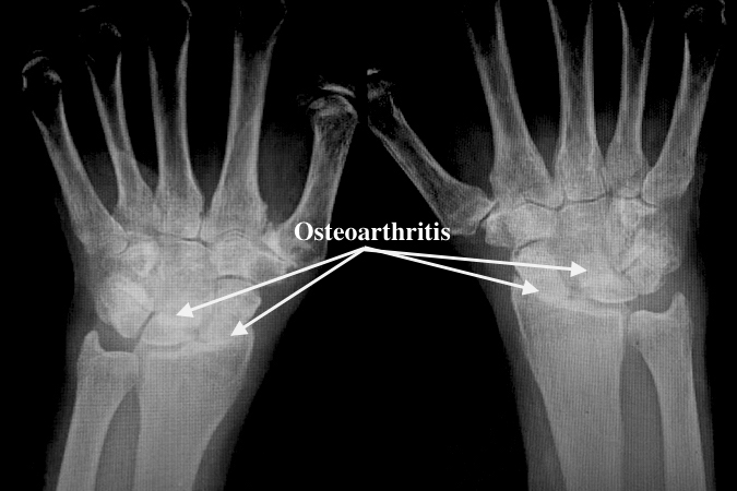
Scapholunate Advanced Collapse (SLAC) is a progressive form of wrist osteoarthritis caused by the chronic disruption of the scapholunate ligament. This instability leads to predictable patterns of carpal collapse, starting with scapholunate dissociation and progressing through stages of arthritis, often resulting in pain, reduced grip strength, and limited wrist motion. Early intervention with physiotherapy can help manage symptoms, slow progression, and improve function.

Scapholunate Advanced Collapse (SLAC): A Physiotherapy Guide

What is Scapholunate Advanced Collapse (SLAC)?

Scapholunate advanced collapse (SLAC) is the most common pattern of wrist arthritis. It is described as a frequently encountered, progressive form of wrist osteoarthritis. SLAC is thought to result from the attenuation or chronic disruption of the scapholunate ligament. This ligament tear leads to altered wrist kinematics, which results in predictable patterns of carpal collapse and subsequent osteoarthritis.

Progression & Affected Joints

SLAC wrist progresses through a predictable sequence of degenerative changes (arthrosis), often described in four stages:

  1. Instability and Dissociation: Disruption of the scapholunate ligament leads to scapholunate dissociation and changes in carpal alignment, such as the lunate assuming an extended posture, potentially causing a dorsal intercalated segment instability (DISI) deformity.
  2. Stage I Arthrosis (Radioscaphoid): Osteoarthritic changes initially occur at the most radial aspect of the radioscaphoid joint, typically manifesting as sharp spurring at the radial side of the scaphoid and at the radial styloid process tip.
  3. Stage II Arthrosis (Entire Radioscaphoid): Degenerative changes progress to involve the entire radioscaphoid joint, followed by narrowing of this articulation.
  4. Stage III Arthrosis (Midcarpal/Capitolunate): Degenerative changes progress to involve the midcarpal joint, specifically the capitolunate joint (and scaphocapitate joint in SLAC wrists).
  5. Stage IV Arthrosis (Advanced): This stage involves advanced pancarpal arthritis. Throughout the progression, the articulation between the lunate and radius (radiolunate joint) is typically preserved until very late in the disease process.

The predictable nature of this progression, where the radiolunate joint is spared until Stage IV, is key to the diagnosis and guides surgical treatment options.

The failure of the scapholunate ligament acts like breaking the hinge on a double door; once the central connection is gone, the two doors (scaphoid and lunate) swing wildly out of alignment, causing them to jam and wear down the surrounding doorframe (radioscaphoid and capitolunate joints) in a predictable pattern, even while the door hinges still connected to the main wall (radiolunate joint) remain functional until the structure entirely collapses.

Causes of SLAC Wrist

The etiology of scapholunate ligament damage can be traumatic or atraumatic.

Traumatic injury can result from a single acute event or multiple repeated events. In a case-control study comparing patients with advanced SLAC wrist (those requiring surgical intervention) to patients with carpometacarpal osteoarthritis (CMC OA), several characteristics were identified as being significantly associated with SLAC wrist:

  • History of Traumatic Injury: Patients with SLAC wrist were significantly more likely to have a history of a traumatic injury to the affected upper extremity (69.5%) compared to the control group (25.9%) (P < .001). This association suggests that injury can be a factor in the development of SLAC wrist. The odds ratio for a history of trauma in SLAC patients was calculated at 6.53.
  • Occupational Load (Manual Labor): SLAC patients were significantly more likely to be involved in a manual labor job (49.0%) compared to the control group (20.0%) (P = .002). Manual labor jobs were defined as professions involving lifting >10 kg intermittently or >2 kg repetitively. The odds ratio for manual labor employment was 3.94.
  • Gender: Patients with SLAC wrist were more likely to be male (80.3%) than the control group (31.1%) (P < .001). The odds ratio for male gender was significantly elevated at 9.03.
  • Age: SLAC patients were significantly younger at the time of surgery (mean 53.1 years) compared to the CMC OA patients (mean 58.3 years) (P = .006). Comparing the estimated age of onset of symptoms (age during treatment minus symptomatic period) showed a 12-year difference (SLAC 42.8 years, CMC OA 54.8 years).

Atraumatic causes leading to the attenuation of the scapholunate ligament and subsequent SLAC wrist include:

  • Calcium pyrophosphate dihydrate (CPPD) crystal deposition disease: Pyrophosphate deposition in the interosseous ligaments (scapholunate) causes ligament laxity and disruption, leading to the rotational alteration of the scaphoid. SLAC wrist is commonly observed in patients with CPPD arthropathy (26% of the time).
  • Rheumatoid arthritis.
  • Neuropathic diseases.
  • β2microglobulin associated amyloid deposition diseases.

Common Symptoms

Symptomatology in SLAC wrist varies significantly, ranging from radiographic findings discovered incidentally in asymptomatic patients to debilitating wrist pain.

Commonly reported findings in symptomatic patients include:

  • Wrist pain (the predominant finding in most presenting cases).
  • Pain can be elicited at the radiocarpal joint and midcarpal joint on physical examination.
  • Dorsoradial swelling.
  • Limited range of wrist motion.
  • Patients undergoing surgical intervention for SLAC wrist often report a prolonged duration of symptoms (mean of 10.3 years).
  • Functional decline, including reduced grip strength, is often reported in those with advanced disease.

To conceptualize the mechanics of SLAC wrist progression, imagine a small wooden raft (the scaphoid) that is crucial for balancing the center boat (the lunate) on the flowing river (the radius). When the strong rope connecting the raft and the center boat (the scapholunate ligament) breaks, the raft becomes unstable and begins to pivot and crash into the riverbank (the radial styloid). This initial, consistent point of impact causes the first signs of wear (Stage I arthritis), eventually wearing down the entire contact area, and finally destabilizing the entire carpal joint structure upstream (midcarpal/capitolunate arthritis, Stages II and III).

Carpal Anatomy and the Scapholunate Ligament

The wrist contains numerous carpal bones, but the pathogenesis of SLAC wrist focuses on the relationship between the scaphoid and the lunate.

  • Scaphoid and Lunate Relationship: The scapholunate interosseous ligament (SLIL) is the primary stabilizing ligament that binds the scaphoid and lunate together. The SLIL is composed of three distinct parts: the dorsal, membranous, and volar components. The dorsal component is the thickest and strongest, making it the most crucial part for stabilization.
  • Secondary Stabilizers: The scaphotrapezium and radioscaphocapitate ligaments are considered secondary stabilizers of the scapholunate articulation and are less vital than the SLIL.

Pathomechanics: The Collapse Process

The pathogenesis of SLAC wrist involves a sequence of anatomical and mechanical changes following the initial injury:

  1. Initial Instability: SLAC wrist is theorized to develop after an inciting event that leads to dissociation of the scaphoid and lunate. The most common mechanism of injury in scapholunate dissociation is trauma causing wrist extension, ulnar deviation, and intercarpal supination.
  2. Loss of Coordinated Motion (Dissociation): When the primary stabilizing ligament (the scapholunate interosseous ligament) is compromised, the scaphoid and lunate lose their synchronous motion and normal alignment.
  3. Malalignment: This instability results in characteristic pathological displacement:

Rotary Subluxation of the Scaphoid: The scaphoid rotates volarly (flexion) and displaces. Its proximal pole often moves dorsally and rotates onto the dorsal aspect of the radius and capitate. This rotary subluxation of the scaphoid is the most common cause of SLAC.

Lunate Extension (DISI): The lunate, unrestrained by the scaphoid, assumes an extended (dorsal) posture, which may result in a Dorsal Intercalated Segment Instability (DISI) deformity.

  1. Abnormal Load Distribution: The altered alignment of the carpus places inordinate stress and load predominantly on the radioscaphoid and capitolunate joints. This is particularly destructive at the radioscaphoid joint due to its elliptical configuration. If the scaphoid rotates (rotary subluxation), this elliptical configuration causes incongruence, shifting the load to the dorsal and volar aspects of the radioscaphoid articulation, leading to destruction and degeneration.
  2. Progressive Arthritis: The mechanical changes lead to a sequential and consistent progressive pattern of degenerative changes (arthrosis - as explained earlier):

SLAC Variants and Related Conditions

Other causes that result in the SLAC pattern of degenerative changes include:

  • Scaphoid nonunion advanced collapse (SNAC): This is due to a non-united fracture of the scaphoid. Although the scapholunate ligament is usually preserved in SNAC (unlike SLAC), it shares a very similar pathophysiology and progression pattern of arthritic change.
  • Idiopathic avascular necrosis of the scaphoid (Preiser disease).
  • Midcarpal instability.
  • Intra-articular fractures involving the radioscaphoid or capitolunate joint.
  • Perilunate dislocation.
  • Kienbock’s disease.

The sources strongly support the idea that initial untreated carpal instability after ligament injury is the primary mechanical driver of SLAC, and that repetitive load and occupational strain may contribute to the progression and severity, as evidenced by the high association between SLAC patients and manual labor jobs and a history of trauma.

Start Your Journey to 

Better Health Today

Recover faster, move better, and feel stronger with expert physiotherapy. Our team is here to guide you every step of the way.

Physiotherapy Is Essential for SLAC Wrist Management and Recovery

Physiotherapy is a cornerstone of managing a Scapholunate Advanced Collapse (SLAC) wrist. Unlike passive treatments that only dull symptoms, active rehabilitation directly addresses the mechanical and functional issues caused by this degenerative and instability-driven condition. Key reasons why physiotherapy is critical include:

  • Improving Load Tolerance and Movement Quality: Exercise-based therapy strengthens the muscles around the wrist, enabling them to absorb shock and support joints during activity. The stronger these muscles are, the more load they can take off the damaged wrist bones. This means better load tolerance (e.g. weight-bearing through the wrist) and smoother, safer movement patterns benefits that painkillers or rest alone cannot provide. Notably, avoiding activity altogether can worsen stiffness and pain over time whereas guided exercise increases blood flow and nutrients to cartilage, helping relieve pain and improve mobility
  • Secondary Stabilizer Strengthening: After scapholunate ligament injury, the body relies on muscular support to stabilize the wrist. Physiotherapy targets the “SL-friendly” muscles such as the flexor carpi radialis (FCR), extensor carpi radialis longus (ECRL), and Abductor Pollicis Longus (APL) which protect the scaphoid from being dragged into a subluxed position away from the lunate. Strengthening these secondary stabilizers can significantly improve wrist stability in the absence of an intact ligament (Therapists may caution against overusing certain muscles like the extensor carpi ulnaris early on, since an unopposed ECU tends to widen the scapholunate gap By also strengthening smaller intrinsic hand muscles and other forearm muscles, therapy increases overall grip strength and support to the collapsing carpal structure
  • Joint Protection via Bracing and Technique Training: A hand therapist can fit patients with custom wrist orthoses (braces) and teach proper alignment strategies for daily tasksThese supports and movement modifications protect the joint surfaces from further wear. For example, learning to keep the wrist in a safer neutral position when pushing up from a chair or doing a push-up can reduce painful shear on arthritic areas. The therapist will educate on “joint protection” techniques ways to lift, carry, or bear weight that minimize stress on the wrist. This proactive training prevents repetitive trauma to the cartilage by ensuring the wrist is braced and aligned during impact or heavy lifting tasks.
  • Maintaining Mobility in Safe Planes: Even though parts of the wrist are arthritic, it’s important to keep the wrist moving in pain-free ranges to prevent stiffness. Therapists focus on planes of motion that do not grind the degenerating joint surfaces. For instance, the dart thrower’s motion (moving the wrist from a radially extended position to ulnarly deviated flexion) primarily uses the midcarpal joint and minimizes stress on the injured scapholunate ligament. Incorporating such wrist motions allows patients to regain range of motion and dexterity in the non-arthritic portions of the wrist while unloading the damaged radiocarpal interface. This means patients can improve wrist flexibility and extension without painfully “point-loading” the bone-on-bone areas.
  • Gradual Return-to-Activity Progressions: Physiotherapy provides a structured pathway to safely resume sports or demanding activities. Rather than abrupt or fearless return (which risks re-injury), the therapist will introduce progressive loading based on the patient’s tolerance. For example, after building baseline strength and stability, patients might progress from light weight-bearing exercises to modified push-ups, and eventually to full impact activities (like boxing or weightlifting) using proper form and bracing. This stepwise approach allows the wrist to adapt and increases capacity for gripping, punching, or pressing tasks over time. In one case, an 8-week program of targeted stabilization and proprioceptive exercises led to improved range of motion and a pain-free return to work for a patient with chronic scapholunate injury.Such outcomes illustrate how active rehab restores functional “kinetic collapse” control essentially retraining the wrist to handle stress without collapsing further.

Passive treatments alone like NSAIDs, corticosteroid creams, rest, or ultrasound may provide temporary symptom relief but do not stop the underlying instability or weakness. Medications and modalities can reduce inflammation or pain perception, yet they fail to retrain the dynamic support of the wrist or prevent progressive collapse. In contrast, physiotherapy directly tackles the root problems: by improving strength, neuromuscular control, and joint mechanics, it slows the degenerative cycle instead of just masking pain. This is why experts emphasize an interprofessional approach (including physical therapy) for SLAC wrists to enhance patient outcomes beyond what pain management alone can achieve

Prognosis: What to Expect with SLAC Wrist Physiotherapy Care

SLAC is a progressive degenerative condition. It results from chronic disruption of the scapholunate ligament leading to predictable patterns of carpal collapse and subsequent osteoarthritis. Because it is degenerative, it is not expected to "heal back to normal anatomy". However, symptoms and decline can be managed and slowed.

Recovery Timeline and Chronic Management

The progression of SLAC can be slow. Patients who eventually require surgical intervention for severe SLAC wrist report a significantly longer duration of symptoms prior to treatment, averaging 10.3 years. This long history supports the idea that the condition requires long-term management. The physiotherapy programme timeline varies based on patient goals, symptom response, and their ability to self-manage, evolving to a chronic management approach.

Typical Expected Outcomes

A service evaluation of the specialized rehabilitation programme for stage one instability showed functional improvements that consistently exceeded minimal clinically important difference (MCID) values.

  1. Pain becomes less frequent, less irritable, and settles quicker after load: The mean Visual Analogue Scale (VAS) pain score reduced by 5.5 points (MCID is 1.37).
  2. Grip and capacity improve: Maximal grip strength (MGS) improved by a mean of 11.5 kg (MCID 6.5 kg). Pain-Free Grip (PFG) improved by a mean of 22 kg. The Grip Strength Ratio (GSR), comparing the injured to the non-injured hand, improved by a mean of 45% (MCID 19%).
  3. Functional measures improve: Mean Quick Disability of Arm, Shoulder and Hand (Quick-DASH) scores, a measure of disability, improved by 33.96% (MCID 14). Mean quality-adjusted life years (QALYs) improved by 0.187 (MCID 0.05).
  4. Return to training and daily tasks becomes safe: Patients who had higher functional demands, such as those in manual work or sports like national level rowing, were successfully progressed through and completed the highest stages of the demanding rehabilitation program.

The conservative management of Scapholunate Advanced Collapse (SLAC) wrist, particularly in its earlier stages where symptoms are manageable without immediate surgery, focuses on stabilizing the carpus through dynamic muscular control and modifying load tolerance. While generalized nonoperative measures typically include wrist immobilization with splints, oral analgesics, and corticosteroid injections, advanced rehabilitation programmes target the underlying instability inherent in the collapse.

The primary evidence base for active rehabilitation focuses on stage one scapholunate instability, the precursor to SLAC, but the principles of restoring dynamic stability are relevant for nonoperative management of mild SLAC arthropathy.

Conservative Physiotherapy Treatment and Exercise Selection for SLAC Wrist

Assessment Priorities

The assessment of SLAC and SNAC wrists requires a multifaceted approach, including a detailed history, physical examination, and radiographic imaging. The following priorities should guide the clinician's evaluation:

1. Grip Dynamometry

Grip strength is an essential functional measure for patients with wrist instability. Both Maximal Grip Strength (MGS) and Pain-Free Grip (PFG) should be assessed using a dynamometer. The Grip Strength Ratio (GSR), which compares the affected to the non-injured hand, helps account for individual factors influencing grip strength. Studies show that improvements in MGS, PFG, and GSR are clinically significant markers of progress in rehabilitation.

2. Wrist Range of Motion (ROM) Assessment

Patients with SLAC often present with reduced wrist range of motion, particularly in extension and ulnar deviation. Measuring Active Range of Motion (AROM) and Passive Range of Motion (PROM) is essential for tracking progress. Maintaining or improving wrist motion, especially at the end of range extension, is critical for rehabilitation and joint function.

3. Symptom Scoring

Pain levels and functional disability should be monitored throughout treatment. The Visual Analogue Scale (VAS) is an effective tool for assessing pain intensity, while the Quick Disability of Arm, Shoulder, and Hand (Quick-DASH) questionnaire provides a measure of functional impairment.

4. Carpal Instability Screening

It is essential to evaluate wrist instability through clinical tests. The Watson test, which assesses pain related to scapholunate instability, and the SL Ballottement test, which detects both pain and laxity, are valuable diagnostic tools. Pain in the radiocarpal and midcarpal joints often accompanies carpal instability.

5. Screening for Red Flags

Clinicians must differentiate wrist pain due to arthritis from other conditions like carpal tunnel syndrome, De Quervain’s tenosynovitis, and flexor carpi radialis (FCR) tendonitis. A thorough examination helps rule out these non-arthritic sources of pain, which could otherwise complicate the diagnosis.

Strengthening Exercises to Support and Stabilize the Scapholunate Interval

The rehabilitation of SLAC wrist focuses on restoring dynamic wrist stability by improving neuromuscular control and addressing the structural changes in the scapholunate ligament. The exercise program should target both the forearm stabilizers and the intrinsic muscles of the hand and wrist.

A. Forearm Stabilizers and Tendon Tolerance

Isometric strengthening exercises are essential to activate the muscles responsible for stabilizing the scaphoid. These exercises are typically performed with resistance bands, starting with moderate intensity and progressing as tolerated. The goal is to restore the muscle balance around the scaphoid and lunate to reduce stress on the scapholunate ligament.

Key Exercise Categories:

  • Scaphoid Supinators: Focused strengthening of the Extensor Carpi Radialis Longus (ECRL), Abductor Pollicis Longus (APL), and Flexor Carpi Radialis (FCR) muscles creates a stabilizing supination moment that improves the alignment of the scaphoid with the lunate. This can prevent further stress on the scapholunate ligament, particularly the dorsal portion. Exercises should begin with the wrist in a supinated position, progressing towards neutral and pronation as strength increases.
  • Tendon Tolerance Progression: Initially, isometric exercises should be performed at different wrist angles (30° increments) to enhance tendon tolerance. As strength improves and symptoms decrease, dynamic strengthening through the full range of motion can be introduced, using resistance bands.
  • Muscles to Avoid: Exercises should avoid over-recruiting muscles that exacerbate scapholunate instability, such as the Extensor Carpi Ulnaris (ECU) and Flexor Carpi Ulnaris (FCU), which may increase the scapholunate gap.

B. Hand and Carpal Intrinsic Activation

At this stage, the focus shifts to proprioceptive rehabilitation, which is designed to improve joint position sense and neuromuscular control. This is critical for maintaining joint stability during functional activities.

Key Exercise Categories:

  • Short-arc Weight-bearing Loading: Early weight-bearing activities, such as bilateral hand placement on a ball (for rolling and circle motions) or wall press-ups, help activate muscles around the wrist while promoting stability. As the patient progresses, exercises can be made more challenging by introducing unstable surfaces (Bosu ball, wobble board) or progressing to four-point kneeling or plank positions.
  • Proprioceptive/Endurance Tools: The use of a Powerball® (gyroscopic device) can help enhance neuromuscular control through multidirectional forces and reactive muscle contractions. For patients who struggle to initiate Powerball exercises, 'auto-start' models may be a helpful alternative.

C. Kinetic Chain Control for High Load Activities

In the final stage of rehabilitation, the focus is on reconditioning the wrist for high-demand activities, such as manual labor or sports. This phase requires the introduction of higher-load and plyometric exercises to prepare the wrist for return to full function.

Key Exercise Categories:

  • High Load Exposure: Incorporating upper limb kinetic chain conditioning is essential for patients with high rehabilitation demands. Early plyometric exercises, like tennis ball drop/catch, can promote explosive movement and dynamic stability. As strength and endurance improve, more advanced exercises such as clap press-ups can be used to prepare the wrist for return to sport.

Progression criteria include achieving pain-free weight-bearing at lower loads before advancing to higher-intensity exercises.

Mobility That Helps Without Aggravating Arthritic Joints

Maintaining mobility is essential, but it must be executed in a way that minimizes stress on the degenerated radioscaphoid and capitolunate joints.

  1. Gentle Flexion/Extension Rocking without Compression Loads (Controlled Movement): The initial focus in therapy is establishing conscious control and isometric loading. When progressing to dynamic exercises, AROM exercises should involve moving the wrist while actively aiming to maintain a neutral deviation. Progression criteria specifically require the patient to maintain control through the range of movement, particularly end of range extension.
  2. Unloading Degenerating Joints: Rehabilitation should preferentially be initiated in supination because forearm pronation has been shown to increase carpal load. This conscious effort helps unload the carpus in the presence of existing arthrosis.

FAQs for SLAC and Physiotherapy

Is SLAC serious, and what is the difference between SLAC and a scapholunate sprain?

  • SLAC is a pattern-based progressive carpal arthritis from old SL instability.
  • SLAC wrist is a serious progressive degenerative condition and the most common cause of osteoarthritis involving the wrist.
  • It is defined as a wrist osteoarthropathy that occurs secondary to altered stress around an unstable scaphoid. It results from the attenuation or chronic disruption of the scapholunate ligament.
  • This chronic instability leads to predictable patterns of collapse, including rotary subluxation of the scaphoid (volar flexion) and the lunate assuming an extended posture (DISI).
  • SLAC wrist follows a sequential progression through up to four stages of degenerative arthrosis.

Difference from Scapholunate Sprain/Instability:

  • Scapholunate instability refers to the loss of synchronous motion between the scaphoid and lunate, often classified in early stages (Stage 1) as a partial scapholunate ligament injury.
  • SLAC wrist is the diagnosis given when the instability has progressed to definite cartilage loss (arthritis), typically beginning at the radioscaphoid joint. Stage 6 SL instability is specifically described as chronic SLIL disruption with cartilage loss.

Can SLAC lead to surgery, and is PT necessary to rebuild movement quality and slow collapse?

  • Yes, SLAC can lead to surgery.
  • SLAC wrist can lead to debilitating wrist pain and functional decline, including limited motion and reduced grip strength.
  • When the degenerative pattern progresses to an advanced pattern of SLAC wrist, surgical intervention is often required.
  • Surgical options are warranted if symptoms persist or worsen after non-surgical treatments (splinting, anti-inflammatories, and injections) are attempted.
  • Surgical procedures for SLAC (Stages II and III) are diverse and include motion-preserving options like Proximal Row Carpectomy (PRC), and partial arthrodesis like four-corner arthrodesis.
  • Physiotherapy is essential because passive treatments alone (splinting, injections) provided only minimal alleviation of pain
  • Active rehabilitation, focusing on dynamic wrist stability, attempts to activate secondary stabilizers like the FCR, ECRL, and APL to create a supination moment on the scaphoid that is protective to the scapholunate ligament area.

Recover from SLAC– Book a Physio Assessment Today

Don’t let wrist pain, weakness, or clicking hold you back from training, work, or everyday tasks. Whether you're lifting, gripping, or returning to sport, our expert physiotherapists are ready to help you stabilize your wrist, protect your joints, and regain lasting strength.

Book Your Specialized Assessment Today:

Team

Expert Insights

Explore the latest articles written by our clinicians