Why It Matters
Entrapment often mimics knee joint pathology, leading to delayed diagnosis unless carefully assessed.
Causes and Risk Factors
- Iatrogenic (Post-Surgical): This is the most common cause. Sensory changes are reported in up to 77% of patients following knee arthroscopy and are also frequent after ACL reconstructions, total knee arthroplasties (TKA), and saphenous vein harvesting for bypass surgery.
- Trauma: Direct blows to the medial thigh or knee, or falls that stretch the leg, can trigger symptoms.
- Overuse: Repetitive friction from running (especially with poor pelvic stability or hip rotator fatigue) can lead to spontaneous entrapment
Why Physiotherapy is Essential
Physiotherapy and targeted rehabilitation are critical for managing saphenous nerve entrapment, particularly when the etiology is non-surgical or related to biomechanical stress. The sources provide specific evidence supporting the techniques and timelines mentioned in your query.
- Nerve Gliding and Neural Tension: Nerve gliding (or flossing) is a cornerstone of conservative treatment, designed to increase nerve excursion and restore normal neurodynamics. Research indicates that nerve gliding can increase nerve excursion by up to 30% compared to tensioning techniques.
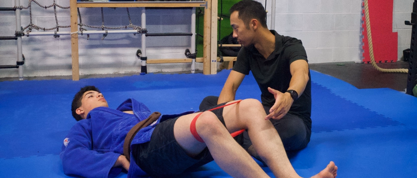
- Manual Therapy and Soft Tissue Release: Techniques such as Myofascial Release and Active Release Technique (ART) are used to manually release the saphenous nerve from entrapment sites, such as the vastoadductor membrane or the adductor canal. These treatments focus on reducing friction and mechanical shear where the nerve pierces fascial structures. Additional modalities may include vibration therapy and acupuncture to address hypertonic musculature and neurogenic pain.
- Biomechanics and Pelvic Stability: Improving biomechanics is vital, as pelvic instability and hip external rotator fatigue are known risk factors that increase valgus loading on the knee, thereby placing tension on the nerve through the adductor canal. Rehabilitative exercises—such as ball squats and single-leg excursions—aim to re-train gluteal activation and multiplanar endurance to reduce this stress.
- Gait and Movement Strategies: Addressing "egg-beater" gait patterns (where the patient crosses midline during running) through gait retraining can prevent the repetitive irritation of the nerve during sport mechanics.
Prognosis: Recovery Timeline
While the specific week-by-week brackets in your query (e.g., "4–6 weeks") are helpful general guidelines, the sources provide documented case-study timelines that vary based on severity:
- Mild/Spontaneous Cases: In one case of a competitive runner, symptoms were fully resolved in one week following just two sessions of Active Release Technique and the initiation of at-home exercises.
- Moderate/Chronic Cases: A patient with a year-long history of pain achieved a 90% recovery after 8 weeks of twice-weekly treatments involving nerve flossing and muscle release. Another case of chronic medial knee pain saw complete resolution within a few weeks after the treatment plan was revised to focus specifically on peripheral nerve entrapment rather than standard bursitis protocols.
- Chronic and Post-Surgical Nuance: The sources note that while many patients respond to conservative care, iatrogenic (post-surgical) cases involving neuromas or severe scarring may be recalcitrant to physical therapy and often require nerve blocks, neurolysis, or neurectomy for relief. Some reports document patients suffering from symptoms for months or even years before an accurate diagnosis is made and appropriate treatment begins
Physiotherapy Treatment Plan
A comprehensive physiotherapy plan for saphenous nerve entrapment focuses on reducing mechanical irritation at entrapment sites like the adductor canal and the vastoadductor membrane while correcting the biomechanical faults that cause nerve tension.
Phase 1: Symptom Relief (0–2 weeks)
- Activity Modification: Avoid activities that stretch the nerve or cause repetitive friction, such as prolonged standing, running, or climbing stairs. In pediatric cases, avoiding lying on the affected side or activities involving hip hyperextension is recommended to prevent exacerbating neural tension.
- Neural Mobilization: Initiate nerve gliding (flossing) to restore neurodynamics and decrease neural edema. Research suggests that "sliders" can increase nerve excursion by up to 30% without the potential irritation caused by high-tension maneuvers.
- Manual and Soft Tissue Therapy: Utilize Active Release Technique (ART) or myofascial release to address the vastoadductor membrane where the nerve pierces the roof of the adductor canal. Clinical cases also support the use of vibration therapy, acupuncture, and muscle release targeting hypertonic muscles like the sartorius, adductor longus, and psoas.
Phase 2: Nerve Mobility & Biomechanics (2–6 weeks)
- Advanced Saphenous Nerve Glides: Transition to specific gliding techniques performed in a side-lying position. The patient moves from hip extension and abduction with slight knee flexion to increased knee flexion while decreasing hip extension to maintain constant neural tension throughout the nerve's anatomical path.
- Addressing Pelvic Instability: Strengthening is essential to correct a positive Trendelenburg’s sign (contralateral pelvic drop), which is often linked to spontaneous entrapment. Focus on the hip external rotators to prevent the valgus knee loading that increases tension on the nerve through the adductor canal.
- Core and Sagittal Plane Stability: Implement ball squats to re-train gluteal activation and limit anterior weight shifting through the quadriceps. Coronal ball contractions (holding a physioball against a wall while standing on one leg) can improve frontal plane stability.
Phase 3: Functional Integration (6–12 weeks)
- Gait Retraining: Correct "egg-beater" gait patterns where the patient crosses the midline while running, as this creates repetitive friction on the nerve. Running on a marked track can help maintain proper alignment.
- Multiplanar Endurance: Use vector exercises (single leg excursions) that incorporate various degrees of hip rotation to increase the endurance and coordination of the hip stabilizing musculature.
- Sport-Specific Loading: Gradually reintroduce sport mechanics, monitoring for the "fullness" or stabbing sensations typical of the condition. In successful case studies, patients were able to return to ultra-marathon training and competitive skating after 2–8 weeks of structured intervention.
Phase 4: Long-Term Maintenance
- Ergonomic and Movement Education: Educate patients on avoiding prolonged pressure on the medial knee and maintaining hip mobility (e.g., addressing hip flexor shortening identified by the Thomas test).
- Screening for Recurrence: Patients should periodically perform prescribed rehab exercises, as follow-ups have shown that even "recovered" athletes may still exhibit mild pelvic drops or valgus motions during fatigue.
- Recognizing PT Limitations: Physical therapists must recognize that post-surgical entrapment or neuromas often respond poorly to conservative care alone and may require referral for nerve blocks, neurolysis, or neurectomy
Prevention & Long-Term Management
Preventing saphenous nerve entrapment and managing it long-term involves a combination of activity modification, biomechanical correction, and vigilant post-surgical monitoring.
Activity Modification and Protection
- Avoid Excessive Kneeling: Clinical history often reveals that kneeling significantly exacerbates saphenous nerve pain. Long-term management includes avoiding aggravating positions and activities that place direct pressure on the medial knee or adductor canal.
- Avoid Repetitive Friction: For athletes, specifically runners, it is important to avoid an "egg-beater" gait pattern (crossing the midline during strides), as this increases repetitive friction on the nerve. Running on a marked track can help maintain proper alignment.
- Environmental Adjustments: In some cases, simple changes like wrapping the knees or using protective strategies during sports can help manage symptoms for those with a history of medial knee trauma.
Maintaining Strength and Biomechanical Stability
- Offloading the Adductor Canal: Maintaining hip and core strength is vital because pelvic instability and hip external rotator fatigue are known risk factors for spontaneous entrapment. Weakness in these areas leads to valgus knee loading, which increases tension on the nerve as it passes through the adductor canal.
- Targeted Exercises:
- Ball Squats: Used to re-train gluteal activation in the sagittal plane and limit the shifting of weight anteriorly through the quadriceps, which can irritate the nerve.
- Coronal Ball Contractions: These focus on frontal plane pelvic stability and eccentric/concentric control of the hip abductors.
- Multiplanar Endurance: "Vector" or single-leg excursion exercises help maintain the coordination of hip stabilizing muscles during complex movements.
- Regular Stretching and Flexibility
- Addressing Muscle Tightness: Excessive tightness in the internal hip rotators and hip flexors can exacerbate nerve entrapment. Regular stretching of the adductors, quadriceps, and psoas is often part of successful recovery protocols.
- Thomas Test Monitoring: Long-term management should include screening for hip flexor shortening, as this can contribute to the biomechanical stress that triggers saphenous neuritis.
Vigilant Monitoring After Trauma or Surgery
- Post-Surgical Awareness: Since iatrogenic (post-surgical) injury is the most common cause of saphenous nerve issues—occurring in up to 77% of arthroscopy patients—monitoring is essential after ACL reconstructions, total knee arthroplasties (TKA), or varicose vein procedures.
- Delayed Symptom Onset: It is critical to monitor for nerve irritation because symptoms may not manifest immediately following a surgical procedure or trauma, which can lead to a confusing clinical presentation later on.
- Chronic Pain Screening: Patients with recalcitrant medial knee pain that has lasted months to years should be screened specifically for nerve entrapment rather than just structural joint issues
FAQs
- Can this heal without surgery?
- Yes, many cases of saphenous nerve entrapment improve significantly with conservative management, particularly those caused by spontaneous entrapment or minor trauma. Clinical evidence shows that:
- Manual Therapy and Rehabilitation: Techniques such as Active Release Technique (ART), myofascial release, and nerve gliding (flossing) can resolve symptoms by releasing the nerve from fascial entrapment and restoring normal neurodynamics. In one instance, a competitive runner achieved full resolution in just two sessions.
- Pharmacological Options: Medications like Gabapentin or Carbamazepine have been used successfully to treat the neurogenic pain associated with saphenous neuralgia.
- Injections: Local injections of anesthetics (like Bupivacaine) and corticosteroids can provide lasting relief, especially in pediatric cases where symptoms were abolished after one or two treatments.
- However, the sources note that iatrogenic (post-surgical) cases—such as those involving neuromas or severe scarring after an ACL reconstruction or total knee arthroplasty—are often uncooperative with physical therapy and may eventually require more invasive intervention.
- Do I need imaging?
- Imaging is usually not the primary tool for diagnosis, which remains largely clinical, but it can be used to confirm the condition or rule out other pathologies.
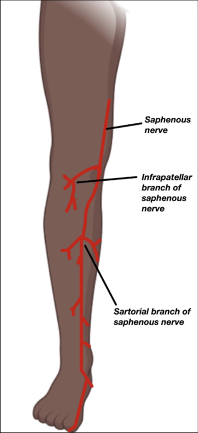
- Clinical Examination: Diagnosis is typically based on subjective history (e.g., burning pain, history of trauma/surgery) and physical tests like a positive Tinel’s sign over the adductor canal or the Reverse Laségue’s test.
- The Gold Standard: Diagnostic nerve blocks are considered the gold standard for confirmation; if an injection of local anesthetic temporarily abolishes the pain, the diagnosis is confirmed.
- Specialized Testing: While MRI and CT scans are sometimes used, they may be normal even when entrapment is present. Somatosensory-evoked potentials (SSEPs) can show increased latency on the affected side, providing electrophysiologic evidence of injury. Musculoskeletal ultrasound is also emerging as a dynamic way to visualize nerve swelling or "flattening" at the entrapment site.
- Will surgery help?
- Surgery is highly effective for refractory or post-surgical cases that do not respond to conservative care.
- Surgical Options: Procedures typically involve neurolysis (releasing the nerve from scar tissue or sutures) or neurectomy (removing a portion of the nerve), especially if a painful neuroma has developed.
- Outcomes: Reported outcomes for surgical interventions are "overwhelmingly positive". Surgery is particularly considered when pain is debilitating and linked to post-operative scarring or nerve transection.
- Ablation: Ablation is the removal or destruction of body tissue or material using methods like heat, cold, laser, or surgery. Less common surgical approaches include cryoablation or radiofrequency ablation to interrupt pain signals
Ready to Relieve Saphenous Nerve Entrapment?
Saphenous nerve entrapment is a frequently overlooked cause of medial knee and leg pain that can mimic common joint injuries. Whether your symptoms stem from previous surgery, trauma, or spontaneous irritation during exercise, our specialized approach is designed to restore your mobility and eliminate neurogenic pain
Our Specialized Approach to Saphenous Nerve Entrapment Rehab
Our comprehensive treatment programs include:
- Detailed Biomechanical Assessment: We evaluate your entire lower limb, looking for pelvic instability, hip external rotator fatigue, and "egg-beater" gait patterns (crossing the midline while running) that increase mechanical shear on the nerve.
- Customized Nerve Mobility Programs: We implement nerve gliding (flossing) techniques designed to increase nerve excursion by up to 30%, restoring neurodynamics without the irritation caused by standard stretching.
- Advanced Manual Therapy for Pain Relief: Our clinicians utilize Active Release Technique (ART®), vibration therapy, and myofascial release to free the nerve from entrapment sites like the adductor canal or the vastoadductor membrane.
- Targeted Strengthening Protocols: We focus on gluteal activation and frontal plane stability using specific tools like ball squats and coronal ball contractions to offload the medial knee.
- Regular Monitoring and Optimization: We track your recovery using validated tools like the Lower Extremity Functional Scale (LEFS) and the Visual Analog Scale (VAS) to ensure your program adjusts as your pain subsides.
Why Choose Our Clinic for Saphenous Nerve Entrapment Treatment?
Evidence-Based Expertise
- Research-Driven Protocols: Our treatments are informed by latest studies on iatrogenic injury (found in up to 77% of post-arthroscopy patients) and spontaneous entrapment in athletes.
- Specialized Neurodynamic Training: We are experts in nerve tension testing, such as the Reverse Laségue’s test, to accurately differentiate nerve pain from medial meniscus tears or MCL injuries.
- Proven Results: We have a track record of successfully returning ultra-marathon runners, competitive skaters, and weightlifters to their peak performance.
Personalized Care
- Precision Diagnosis: We utilize palpatory Tinel’s testing at key anatomical landmarks—like the exit of Hunter’s canal—to pinpoint the exact site of irritation.
- Individualized Plans: We recognize that pediatric cases may present atypically with referred abdominal pain, while post-surgical cases may require a more cautious approach.
- Collaborative Referrals: If conservative care isn't enough, we coordinate with specialists for gold-standard diagnostic nerve blocks or surgical options like neurolysis.
Comprehensive Recovery Support
- Daily Activity Management: We provide education on avoiding aggravating factors, such as kneeling, deep squats, or prolonged standing.
- Safe Return to Sport: We offer specific guidance on gait retraining and multiplanar endurance to ensure your return to running or cutting sports is permanent.
- Long-Term Prevention: Our programs include ongoing screening for hip flexor shortening and muscle imbalances to prevent the recurrence of entrapment
Take the First Step Toward Recovery
Don't let saphenous nerve entrapment limit your activities or affect your daily life. Our experienced team is ready to help you build a strong foundation for lasting recovery.
Book Your Specialized Assessment Today:
Phone: 905-669-1221
Location: 398 Steeles Ave W #201, Thornhill, ON L4J 6X3
Online Booking: www.vaughanphysiotherapy.com
Serving communities across Thornhill, Langstaff, Newtonbrook, Willowdale, North York, Markham, Richmond Hill, Concord, and North Toronto.
Conveniently located in the heart of Thornhill, offering flexible scheduling to accommodate your recovery needs.
Created by Sara Lam

.webp)
.webp)
.webp)


.avif)


