

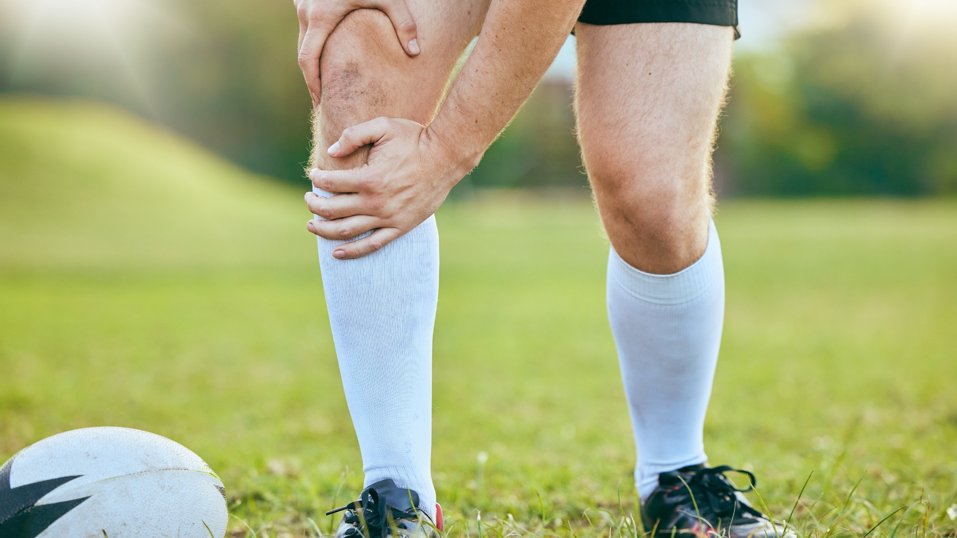

Runner kneeling on the road holding a painful knee after a kneecap injury

Patellar Subluxation/Dislocation

The kneecap slips partly (subluxation) or fully (dislocation) out of its groove, usually toward the outside of the knee, after a twist or pivot.

Did your kneecap suddenly shift out of place while you were pivoting, landing, or changing direction? Did your knee give way, swell up, and leave you nervous to trust it again? If so, you may have had a patellar subluxation or dislocation, a common knee injury in teens and young athletes.

At Vaughan Physiotherapy Clinic, we help athletes, students, and active adults recover after the kneecap has slipped out of place, whether it happened once or keeps happening. The goal is not just to calm the swelling, but to rebuild the strength and control that keep the kneecap where it belongs, and to work out whether you need a surgical opinion.

Here is what patellar subluxation and dislocation are, how to recognize them, what they can be confused with, and what the research says about getting back to sport and daily life.

What Is Patellar Subluxation/Dislocation?

Your patella (kneecap) sits in a shallow groove at the front of the thigh bone called the trochlea. When the kneecap is forced out of that groove, it almost always shifts toward the outside of the knee. This is called a lateral patellar dislocation.

  • Subluxation: the kneecap slides partly out of the groove and pops back on its own. People often describe the knee "giving way" or "shifting"
  • Dislocation: the kneecap comes fully out of the groove. It often slides back in when the knee is straightened, but sometimes it stays out and needs to be put back by a medical professional

In a large population study from the United States, first-time kneecap dislocation happened at a rate of about 23 per 100,000 people each year. The rate was far higher in teenagers aged 14 to 18, and it was similar in males and females (Sanders et al., 2018).

First-Time vs Recurrent Instability

Clinicians usually separate a first-time (primary) dislocation from recurrent instability, where the kneecap keeps slipping out. This matters because the treatment approach and the chance of surgery are different. Across studies of non-surgical care, roughly 3 in 10 people have another dislocation (Moiz et al., 2018), though the risk ranges widely depending on age and knee shape (Frings et al., 2020).

Understanding the Anatomy

Several structures work together to keep the kneecap on track:

  • Trochlear groove: the channel on the thigh bone that the kneecap glides in. A shallow or flat groove (trochlear dysplasia) gives the kneecap less to hold on to
  • Medial patellofemoral ligament (MPFL): a band on the inner side of the knee that acts as the main restraint against the kneecap sliding outward. On MRI, it is torn in about 98 percent of people after a dislocation (Migliorini et al., 2021)
  • Quadriceps muscles: the large thigh muscles that pull on the kneecap and control the knee during landing and cutting
  • Patellar tendon and tibial tubercle: the tendon below the kneecap and the bony bump it attaches to. If the bump sits further to the outside, or the kneecap rides high (patella alta), the kneecap is more likely to slip
  • Cartilage: the smooth lining under the kneecap and in the groove, which can be bruised or chipped when the kneecap dislocates and relocates

Recognizing the Symptoms

A dislocation usually happens with a twist or pivot on a bent knee, often with the foot planted, or from a direct blow. Common signs include:

  • A sudden shift, pop, or "knee went out" feeling, often followed by falling
  • A visible bump on the outside of the knee if the kneecap stays out
  • Rapid swelling within hours, sometimes from bleeding inside the joint
  • Pain and tenderness on the inner side of the kneecap, where the MPFL tears
  • Difficulty straightening the knee or putting full weight on it
  • A feeling of apprehension: worry that the knee will give way again, especially when someone pushes the kneecap outward

With a subluxation, symptoms are usually milder: a brief shift, some pain, and a knee that feels unreliable afterward.

What Makes It Worse?

  • Young age and open growth plates: children and younger teens have a higher chance of the kneecap dislocating again (Frings et al., 2020)
  • Knee shape: a shallow trochlear groove, a high-riding kneecap, or a tibial tubercle that sits further to the outside all raise the risk, and the risk climbs further when several of these are present together (Frings et al., 2020)
  • Previous dislocation on the other side: this roughly triples the risk of another dislocation in some studies (Frings et al., 2020)
  • General joint looseness and family history: both are linked with kneecap instability (Migliorini et al., 2021)
  • Pivoting and cutting sports: basketball, soccer, dance, gymnastics, cheer, and similar activities put the kneecap under twisting load
  • Weak or poorly controlled hip and thigh muscles: which can let the knee collapse inward during landing and turning

What Else Could It Be?

A twisting knee injury with swelling can have several causes, and some need very different treatment. Part of our assessment is ruling these in or out:

  • Anterior cruciate ligament (ACL) tear: also causes a pop, giving way, and fast swelling after a pivot. The two injuries can feel very similar in the first few days
  • Meniscus tear: pain along the joint line, catching, or locking
  • Osteochondral fracture or loose body: a piece of cartilage or bone knocked off during the dislocation. Loose fragments show up in about 1 in 9 people on imaging after a dislocation (Migliorini et al., 2021)
  • Medial collateral ligament (MCL) sprain: pain on the inner side of the knee after a sideways force
  • Patellar fracture or quadriceps or patellar tendon rupture: difficulty or inability to lift the straight leg
  • Patellofemoral pain: aching around the kneecap with stairs, squatting, or sitting, without true episodes of the kneecap slipping out

When to See a Doctor First

Go to an emergency department or see your physician promptly if you have:

  • A kneecap that is still out of place. Do not try to force it back yourself
  • Large, fast swelling within the first few hours, or a knee that feels tight and full, which may mean bleeding in the joint or a fracture
  • A knee that is locked and will not straighten, which may point to a loose fragment or meniscus tear
  • Inability to lift your straight leg off the bed or to put any weight on the leg
  • Numbness, tingling, or a cold or pale foot after the injury
  • A hot, red, swollen knee with fever, especially without a clear injury

After a first dislocation, an X-ray is usually needed, and an MRI is often recommended to check the ligament and cartilage (Migliorini et al., 2021). Our physiotherapists screen for these signs at your first visit and will coordinate with your physician if imaging or a referral to a knee specialist is needed.

Related reading: Front of knee pain and kneecap problems are also covered in our guides to patellofemoral pain syndrome, chondromalacia patella and hypermobility.

Start Your Journey to 

‍Better Health Today

Recover faster, move better, and feel stronger with expert physiotherapy. Our team is here to guide you every step of the way.

Why Physiotherapy Is Essential

After the kneecap slips out, the knee is left swollen, sore, and weak, and the thigh muscles often switch off. Many people also lose trust in the knee. Rest alone may settle the pain, but it does not rebuild the strength, control, and confidence that help protect the knee when you return to sport.

For most first-time dislocations, non-surgical care built around rehabilitation is the usual starting point. A Cochrane review of 10 trials (519 people) found that surgery lowered the chance of another dislocation, but it was uncertain whether surgery led to better function, and surgery may bring more complications in the first two years. The overall quality of that evidence was very low (Smith et al., 2023). A systematic review of non-surgical care found that early, active rehabilitation is justified, even though no single program has been proven better than another (Moiz et al., 2018).

Physiotherapy addresses the main drivers:

  • Swelling and pain: settling the joint so the thigh muscles can work again
  • Range of motion: restoring full bending and straightening without provoking the kneecap
  • Strength: rebuilding the quadriceps, hips, and calves, which help control the knee during landing and turning
  • Control and confidence: balance, landing, and change-of-direction training so the knee feels reliable again
  • Risk screening: identifying the people whose knee shape or history puts them at higher risk, so they can see a surgeon early

Your Treatment Plan at Vaughan Physiotherapy Clinic

Phase 1: Assessment and Protection (Weeks 0 to 2)

Your first visit focuses on understanding the injury and your risk of it happening again. A full assessment includes:

  • History: how the injury happened, whether the kneecap stayed out, how fast the knee swelled, and whether you or a family member have had kneecap problems before
  • Swelling and range of motion: how much fluid is in the joint and how far the knee bends and straightens
  • Tenderness and ligament checks: feeling along the inner side of the kneecap where the MPFL tears, and gently testing the ACL, MCL, and meniscus
  • Kneecap position and apprehension: how the kneecap sits and moves, and whether gentle pressure outward makes you feel it will slip
  • Hip, ankle, and general joint looseness: factors that change how load passes through the knee
  • Imaging review: X-ray and MRI results if you have them, including signs of a shallow groove, a high-riding kneecap, or a loose fragment

Several features predict a higher chance of the kneecap dislocating again, including young age, open growth plates, a shallow trochlear groove, a high-riding kneecap, and a previous dislocation on the other knee. Risk rises with each added factor, and in some groups more than half of people dislocate again (Frings et al., 2020). If your profile looks high risk, or imaging shows a loose piece of bone or cartilage, we will recommend an early opinion from an orthopaedic surgeon.

Early care usually includes:

  • Swelling control: elevation, compression, and gentle muscle pumping exercises
  • Protected walking: crutches for a short time if needed, progressing to full weight as comfort allows
  • Short-term support: a brace or taping for comfort and confidence. Studies of braces, taping, and casting after dislocation are small and hard to compare, so there is no clear best option (Moiz et al., 2018)
  • Quadriceps activation: tightening the thigh muscle and straight leg raises to stop it from shutting down
  • Gentle range of motion: working back toward a straight knee and comfortable bending

Phase 2: Restoring Motion and Early Strength (Weeks 2 to 6)

Once swelling is settling, the focus moves to full movement and building a base of strength. Rehabilitation after a kneecap dislocation should target pain and swelling control, restoring motion, and progressive strengthening of both the hip and the knee (Watson et al., 2022).

  • Range of motion: stationary cycling and heel slides to regain full bending
  • Quadriceps strengthening: mini squats, step-ups, and leg press within a comfortable range
  • Hip strengthening: side-lying leg lifts, bridges, and band walks
  • Balance: single-leg standing, progressing to unstable surfaces

Many programs used to focus on one part of the thigh muscle (the vastus medialis oblique, or VMO). In a trial comparing VMO-focused exercise with general quadriceps exercise after a first dislocation, there was no clinically meaningful difference between the two (Moiz et al., 2018). We focus on overall leg strength rather than trying to isolate one muscle.

Phase 3: Building Strength and Control (Weeks 6 to 12)

Hip and knee strengthening
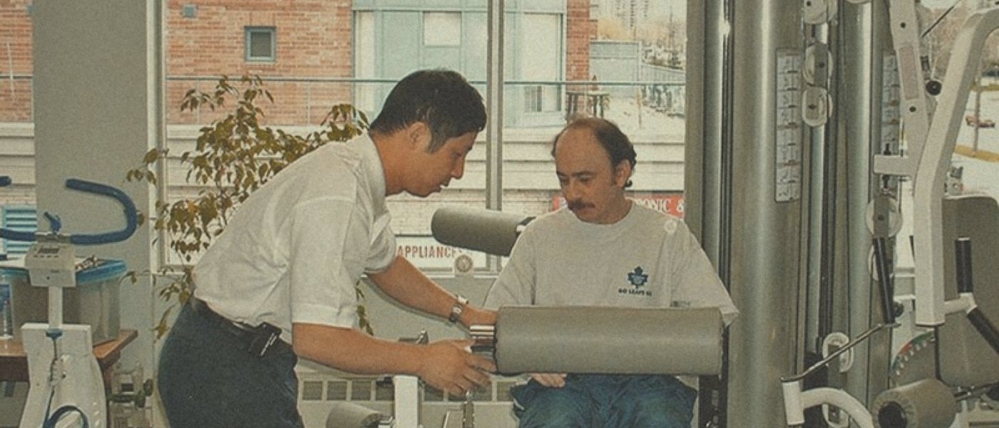
Physiotherapist adjusting a leg extension machine for a patient doing quadriceps strengthening
Quadriceps and hip strengthening help rebuild control of the kneecap after a dislocation.

This phase builds the strength and control your knee needs for sport and daily life. Programs should include both single-joint exercises and whole-leg movements (Watson et al., 2022). In people with kneecap pain, combined hip and knee exercise gives better results than knee exercise alone (Willy et al., 2019). That evidence comes from a related kneecap condition, not dislocation, but the principle fits well. Your program may include:

  • Split squats and lunges, progressing in depth and load
  • Single-leg squats and step-downs with good knee alignment
  • Leg press and knee extension through a safe range
  • Side planks, hip abduction, and hip rotation exercises
  • Calf raises, progressing to single-leg

Neuromuscular and landing training

  • Balance and reaching drills on one leg
  • Two-foot jumping and landing, progressing to single-leg hops
  • Training the knee to stay in line over the foot during landing and cutting

Phase 4: Return to Sport and Full Activity (Week 12+)

  • Running progression: straight-line running first, then curves and changes of pace
  • Agility and cutting drills: gradually adding pivoting and sport-specific movements
  • Plyometrics: hopping, bounding, and side-to-side jumps
  • Return-to-sport testing: before clearance, we compare strength and hop tests between the two legs and use questionnaires about how stable and capable your knee feels (Watson et al., 2022). Decisions should be based on meeting criteria, not just time, and should take into account how mentally ready you feel and what your sport demands (Lampros & Tanaka, 2022)

Beyond Physiotherapy: Complementary Care for Patellar Subluxation/Dislocation

Strengthening, control training, and graded return to sport form the core of care after a kneecap dislocation. Some patients also benefit from other services at our clinic to ease pain and muscle tightness so they can stay consistent with rehab. Here is an honest summary of what the evidence does and does not show for each.

How Acupuncture and Dry Needling Can Help

Acupuncture and dry needling use fine needles to reduce pain and ease tight, tender muscles.

  • What the evidence shows: we did not find any published trials of acupuncture or dry needling for kneecap dislocation or instability. The closest evidence comes from kneecap pain, where the clinical practice guideline advises against dry needling and says acupuncture may be used for pain with caution, since it is not clear that it works better than placebo (Willy et al., 2019)
  • Where it fits: as an optional, short-term aid for pain or thigh and hip muscle tension. It does not stabilize the kneecap
  • Safety: generally very safe. Minor bruising or soreness at needle sites is the most common side effect. We avoid needling over a fresh, swollen joint

How Massage Therapy Can Help

Registered massage therapy focuses on the soft tissues of the thigh, hip, and calf.

  • What the evidence shows: there are no published trials of massage for kneecap dislocation or instability
  • Where it fits: it may help with comfort and with tight thigh and hip muscles during the strengthening phases. We treat it as a supportive add-on, not a stand-alone treatment
  • Safety: very safe. Deep work directly over the injured inner side of the knee is avoided in the early weeks

How Chiropractic and Joint Manipulation Can Help

Chiropractic care uses joint mobilization and manipulation, usually of the spine, hip, knee, and ankle.

  • What the evidence shows: we did not find peer-reviewed trials of chiropractic care or joint manipulation for kneecap dislocation. In kneecap pain, guidelines advise against using spinal, knee, or kneecap manipulation on its own, though gentle kneecap mobilization may be combined with exercise (Willy et al., 2019)
  • Where it fits: for stiffness in the hip, ankle, or back that may affect how you move, always combined with exercise
  • Safety: forceful techniques that push the kneecap outward should be avoided in an unstable knee. Manipulation is not appropriate over a suspected fracture or loose body

Whichever combination you and your clinician choose, these treatments share one purpose: settling symptoms so you can keep training while the real fix, a stronger, better controlled leg, takes hold.

When Is Surgery Considered?

Surgery is usually considered when:

  • A loose piece of bone or cartilage has broken off and needs to be fixed or removed
  • The kneecap keeps dislocating despite a good rehabilitation program
  • Several high-risk features are present after a first dislocation, such as a shallow groove, a high-riding kneecap, and open growth plates. Experts suggest these patients may be considered for earlier surgery to protect the cartilage over the long term (Frings et al., 2020)

The most common operation is MPFL reconstruction, which rebuilds the torn ligament with a tendon graft. Sometimes it is combined with bone procedures that change the groove or the position of the tibial tubercle. In a review of 27 studies, about 85 percent of athletes returned to sport after MPFL reconstruction, but only about 68 percent returned to their previous level, and return took about 7 months on average (Manjunath et al., 2021). If you have surgery, physiotherapy follows your surgeon's protocol and restrictions.

How Long Will It Take?

Recovery depends on how severe the injury was, whether there is cartilage damage, your age and knee shape, and what you are returning to. Most people improve a great deal with non-surgical care, but on average, function does not fully return to normal in every case, and a minority have another dislocation (Moiz et al., 2018). There is little good data on exactly how long non-surgical recovery takes.

In our experience, many people are walking comfortably within a few weeks, with return to pivoting sport taking a few months, and only once strength and hop tests show the injured leg has caught up. After MPFL reconstruction, return to sport takes about 7 months on average (Manjunath et al., 2021).

Prevention and Long-Term Management

  • Keep up your strength work: continue hip and thigh exercises two to three times a week, even after you are back to sport
  • Train landing and cutting: include balance and landing drills in your warm-up
  • Return gradually: build up training load step by step rather than jumping straight back to full games
  • Use support if it helps: a brace or tape can add confidence during sport, but it does not replace strength training
  • Act early: if the knee starts to feel unstable again, book a reassessment rather than waiting for another dislocation
  • Know your risk: if you have a shallow groove, a high-riding kneecap, or problems on both sides, ask about a surgical opinion if episodes continue

Frequently Asked Questions

Is a kneecap dislocation serious?

It is a significant injury, but most people recover well. The main concerns are damage to the cartilage and the chance of it happening again. Repeated dislocations can damage the cartilage under the kneecap over time (Frings et al., 2020), which is why proper rehab and, in some cases, a surgical opinion matter.

Will my kneecap dislocate again?

It might. Across studies of non-surgical care, about 3 in 10 people dislocate again (Moiz et al., 2018). Your own risk depends on your age, knee shape, and history. Young people with several risk factors have a much higher chance, while fully grown people without risk factors have a much lower one (Frings et al., 2020).

Do I need surgery after my first dislocation?

Usually not. Most first-time dislocations are treated without surgery. Surgery is considered if there is a loose fragment, repeated dislocations, or a high-risk profile. Research has not clearly shown that surgery gives better function after a first dislocation, although it does lower the chance of another one (Smith et al., 2023).

Do I need an MRI?

Often, yes. Cartilage and ligament injuries are common after a dislocation, and some researchers recommend MRI for everyone after a dislocation (Migliorini et al., 2021). Your physician or surgeon will decide, and we can help coordinate the referral.

When can I go back to sport?

When you meet the criteria, not on a set date. That means full movement, no swelling, strength and hop tests close to your other leg, and feeling confident in the knee (Watson et al., 2022). For many people after non-surgical care, this takes a few months.

Can acupuncture, massage, or chiropractic replace exercise after a kneecap dislocation?

No. Strengthening and control training are the foundation of recovery. Needling, massage, and joint mobilization have not been tested for this injury and are best used as short-term supports to ease pain and tightness while you rebuild.

Our Specialized Approach at Vaughan Physiotherapy Clinic

Risk-focused assessment: we look at your age, knee shape, and history to judge the chance of it happening again

Early swelling and strength care: getting the thigh muscles working again from the first week

Hip and knee strengthening: a progressive program targeting the whole leg, not just one muscle

Criteria-based return to sport: strength and hop testing before you go back to pivoting and cutting

Integrated care: physiotherapy, massage therapy, acupuncture, and chiropractic under one roof when they add value

Safety first: screening for fractures, loose fragments, and other ligament injuries, with physician or surgeon referral when needed

Take the First Step Toward a Stable, Confident Knee

A kneecap that slips out does not have to end your season or keep you from the activities you love. Our team at Vaughan Physiotherapy Clinic helps patients from Thornhill, Vaughan, North York, Richmond Hill, and surrounding communities rebuild strength and confidence with evidence-based, hands-on care.

Book your assessment today:

Phone: 905-669-1221

Location: 398 Steeles Ave W #201, Thornhill, ON L4J 6X3

Online Booking: www.vaughanphysiotherapy.com

References

Frings J, Balcarek P, Tscholl P, Liebensteiner M, Dirisamer F, Koenen P. Conservative versus surgical treatment for primary patellar dislocation: a systematic review to guide risk stratification. Deutsches Ärzteblatt International. 2020;117(16):279-286. doi:10.3238/arztebl.2020.0279

Lampros RE, Tanaka MJ. Return to play considerations after patellar instability. Current Reviews in Musculoskeletal Medicine. 2022;15:597-605. doi:10.1007/s12178-022-09792-1

Manjunath AK, Hurley ET, Jazrawi LM, Strauss EJ. Return to play after medial patellofemoral ligament reconstruction: a systematic review. American Journal of Sports Medicine. 2021;49(4):1094-1100. doi:10.1177/0363546520947044

Migliorini F, Marsilio E, Cuozzo F, Oliva F, Eschweiler J, Hildebrand F, Maffulli N. Chondral and soft tissue injuries associated to acute patellar dislocation: a systematic review. Life. 2021;11(12):1360. doi:10.3390/life11121360

Moiz M, Smith N, Smith TO, Chawla A, Thompson P, Metcalfe A. Clinical outcomes after the nonoperative management of lateral patellar dislocations: a systematic review. Orthopaedic Journal of Sports Medicine. 2018;6(6):2325967118766275. doi:10.1177/2325967118766275

Sanders TL, Pareek A, Hewett TE, Stuart MJ, Dahm DL, Krych AJ. Incidence of first-time lateral patellar dislocation: a 21-year population-based study. Sports Health. 2018;10(2):146-151. doi:10.1177/1941738117725055

Smith TO, Gaukroger A, Metcalfe A, Hing CB. Surgical versus non-surgical interventions for treating patellar dislocation. Cochrane Database of Systematic Reviews. 2023;(1):CD008106. doi:10.1002/14651858.CD008106.pub4

Watson R, Sullivan B, Stone AV, Jacobs C, Malone T, Heebner N, Noehren B. Lateral patellar dislocation: a critical review and update of evidence-based rehabilitation practice guidelines and expected outcomes. JBJS Reviews. 2022;10(5):e2100159. doi:10.2106/JBJS.RVW.21.00159

Willy RW, Hoglund LT, Barton CJ, Bolgla LA, Scalzitti DA, Logerstedt DS, Lynch AD, Snyder-Mackler L, McDonough CM. Patellofemoral pain: clinical practice guidelines linked to the International Classification of Functioning, Disability and Health from the Academy of Orthopaedic Physical Therapy of the American Physical Therapy Association. Journal of Orthopaedic & Sports Physical Therapy. 2019;49(9):CPG1-CPG95. doi:10.2519/jospt.2019.0302

Disclaimer: This article is for educational purposes only and does not replace professional medical advice. A kneecap that has slipped out of place can be linked with fractures, cartilage damage, and other ligament injuries that need medical assessment and imaging. If your kneecap is still out of place, or your knee is locked, rapidly swelling, or unable to bear weight, seek urgent medical care. Always consult a qualified healthcare provider for diagnosis and treatment specific to your condition.


Team

Condition guide

Patellar Subluxation/Dislocation

The kneecap slips partly (subluxation) or fully (dislocation) out of its groove, usually toward the outside of the knee, after a twist or pivot.

Runner kneeling on the road holding a painful knee after a kneecap injuryPatellar Subluxation/Dislocation

Did your kneecap suddenly shift out of place while you were pivoting, landing, or changing direction? Did your knee give way, swell up, and leave you nervous to trust it again? If so, you may have had a patellar subluxation or dislocation, a common knee injury in teens and young athletes.

At Vaughan Physiotherapy Clinic, we help athletes, students, and active adults recover after the kneecap has slipped out of place, whether it happened once or keeps happening. The goal is not just to calm the swelling, but to rebuild the strength and control that keep the kneecap where it belongs, and to work out whether you need a surgical opinion.

Here is what patellar subluxation and dislocation are, how to recognize them, what they can be confused with, and what the research says about getting back to sport and daily life.

What Is Patellar Subluxation/Dislocation?

Your patella (kneecap) sits in a shallow groove at the front of the thigh bone called the trochlea. When the kneecap is forced out of that groove, it almost always shifts toward the outside of the knee. This is called a lateral patellar dislocation.

  • Subluxation: the kneecap slides partly out of the groove and pops back on its own. People often describe the knee "giving way" or "shifting"
  • Dislocation: the kneecap comes fully out of the groove. It often slides back in when the knee is straightened, but sometimes it stays out and needs to be put back by a medical professional

In a large population study from the United States, first-time kneecap dislocation happened at a rate of about 23 per 100,000 people each year. The rate was far higher in teenagers aged 14 to 18, and it was similar in males and females (Sanders et al., 2018).

First-Time vs Recurrent Instability

Clinicians usually separate a first-time (primary) dislocation from recurrent instability, where the kneecap keeps slipping out. This matters because the treatment approach and the chance of surgery are different. Across studies of non-surgical care, roughly 3 in 10 people have another dislocation (Moiz et al., 2018), though the risk ranges widely depending on age and knee shape (Frings et al., 2020).

Understanding the Anatomy

Several structures work together to keep the kneecap on track:

  • Trochlear groove: the channel on the thigh bone that the kneecap glides in. A shallow or flat groove (trochlear dysplasia) gives the kneecap less to hold on to
  • Medial patellofemoral ligament (MPFL): a band on the inner side of the knee that acts as the main restraint against the kneecap sliding outward. On MRI, it is torn in about 98 percent of people after a dislocation (Migliorini et al., 2021)
  • Quadriceps muscles: the large thigh muscles that pull on the kneecap and control the knee during landing and cutting
  • Patellar tendon and tibial tubercle: the tendon below the kneecap and the bony bump it attaches to. If the bump sits further to the outside, or the kneecap rides high (patella alta), the kneecap is more likely to slip
  • Cartilage: the smooth lining under the kneecap and in the groove, which can be bruised or chipped when the kneecap dislocates and relocates

Recognizing the Symptoms

A dislocation usually happens with a twist or pivot on a bent knee, often with the foot planted, or from a direct blow. Common signs include:

  • A sudden shift, pop, or "knee went out" feeling, often followed by falling
  • A visible bump on the outside of the knee if the kneecap stays out
  • Rapid swelling within hours, sometimes from bleeding inside the joint
  • Pain and tenderness on the inner side of the kneecap, where the MPFL tears
  • Difficulty straightening the knee or putting full weight on it
  • A feeling of apprehension: worry that the knee will give way again, especially when someone pushes the kneecap outward

With a subluxation, symptoms are usually milder: a brief shift, some pain, and a knee that feels unreliable afterward.

What Makes It Worse?

  • Young age and open growth plates: children and younger teens have a higher chance of the kneecap dislocating again (Frings et al., 2020)
  • Knee shape: a shallow trochlear groove, a high-riding kneecap, or a tibial tubercle that sits further to the outside all raise the risk, and the risk climbs further when several of these are present together (Frings et al., 2020)
  • Previous dislocation on the other side: this roughly triples the risk of another dislocation in some studies (Frings et al., 2020)
  • General joint looseness and family history: both are linked with kneecap instability (Migliorini et al., 2021)
  • Pivoting and cutting sports: basketball, soccer, dance, gymnastics, cheer, and similar activities put the kneecap under twisting load
  • Weak or poorly controlled hip and thigh muscles: which can let the knee collapse inward during landing and turning

What Else Could It Be?

A twisting knee injury with swelling can have several causes, and some need very different treatment. Part of our assessment is ruling these in or out:

  • Anterior cruciate ligament (ACL) tear: also causes a pop, giving way, and fast swelling after a pivot. The two injuries can feel very similar in the first few days
  • Meniscus tear: pain along the joint line, catching, or locking
  • Osteochondral fracture or loose body: a piece of cartilage or bone knocked off during the dislocation. Loose fragments show up in about 1 in 9 people on imaging after a dislocation (Migliorini et al., 2021)
  • Medial collateral ligament (MCL) sprain: pain on the inner side of the knee after a sideways force
  • Patellar fracture or quadriceps or patellar tendon rupture: difficulty or inability to lift the straight leg
  • Patellofemoral pain: aching around the kneecap with stairs, squatting, or sitting, without true episodes of the kneecap slipping out

When to See a Doctor First

Go to an emergency department or see your physician promptly if you have:

  • A kneecap that is still out of place. Do not try to force it back yourself
  • Large, fast swelling within the first few hours, or a knee that feels tight and full, which may mean bleeding in the joint or a fracture
  • A knee that is locked and will not straighten, which may point to a loose fragment or meniscus tear
  • Inability to lift your straight leg off the bed or to put any weight on the leg
  • Numbness, tingling, or a cold or pale foot after the injury
  • A hot, red, swollen knee with fever, especially without a clear injury

After a first dislocation, an X-ray is usually needed, and an MRI is often recommended to check the ligament and cartilage (Migliorini et al., 2021). Our physiotherapists screen for these signs at your first visit and will coordinate with your physician if imaging or a referral to a knee specialist is needed.

Related reading: Front of knee pain and kneecap problems are also covered in our guides to patellofemoral pain syndrome, chondromalacia patella and hypermobility.

Why Physiotherapy Is Essential

After the kneecap slips out, the knee is left swollen, sore, and weak, and the thigh muscles often switch off. Many people also lose trust in the knee. Rest alone may settle the pain, but it does not rebuild the strength, control, and confidence that help protect the knee when you return to sport.

For most first-time dislocations, non-surgical care built around rehabilitation is the usual starting point. A Cochrane review of 10 trials (519 people) found that surgery lowered the chance of another dislocation, but it was uncertain whether surgery led to better function, and surgery may bring more complications in the first two years. The overall quality of that evidence was very low (Smith et al., 2023). A systematic review of non-surgical care found that early, active rehabilitation is justified, even though no single program has been proven better than another (Moiz et al., 2018).

Physiotherapy addresses the main drivers:

  • Swelling and pain: settling the joint so the thigh muscles can work again
  • Range of motion: restoring full bending and straightening without provoking the kneecap
  • Strength: rebuilding the quadriceps, hips, and calves, which help control the knee during landing and turning
  • Control and confidence: balance, landing, and change-of-direction training so the knee feels reliable again
  • Risk screening: identifying the people whose knee shape or history puts them at higher risk, so they can see a surgeon early

Your Treatment Plan at Vaughan Physiotherapy Clinic

Phase 1: Assessment and Protection (Weeks 0 to 2)

Your first visit focuses on understanding the injury and your risk of it happening again. A full assessment includes:

  • History: how the injury happened, whether the kneecap stayed out, how fast the knee swelled, and whether you or a family member have had kneecap problems before
  • Swelling and range of motion: how much fluid is in the joint and how far the knee bends and straightens
  • Tenderness and ligament checks: feeling along the inner side of the kneecap where the MPFL tears, and gently testing the ACL, MCL, and meniscus
  • Kneecap position and apprehension: how the kneecap sits and moves, and whether gentle pressure outward makes you feel it will slip
  • Hip, ankle, and general joint looseness: factors that change how load passes through the knee
  • Imaging review: X-ray and MRI results if you have them, including signs of a shallow groove, a high-riding kneecap, or a loose fragment

Several features predict a higher chance of the kneecap dislocating again, including young age, open growth plates, a shallow trochlear groove, a high-riding kneecap, and a previous dislocation on the other knee. Risk rises with each added factor, and in some groups more than half of people dislocate again (Frings et al., 2020). If your profile looks high risk, or imaging shows a loose piece of bone or cartilage, we will recommend an early opinion from an orthopaedic surgeon.

Early care usually includes:

  • Swelling control: elevation, compression, and gentle muscle pumping exercises
  • Protected walking: crutches for a short time if needed, progressing to full weight as comfort allows
  • Short-term support: a brace or taping for comfort and confidence. Studies of braces, taping, and casting after dislocation are small and hard to compare, so there is no clear best option (Moiz et al., 2018)
  • Quadriceps activation: tightening the thigh muscle and straight leg raises to stop it from shutting down
  • Gentle range of motion: working back toward a straight knee and comfortable bending

Phase 2: Restoring Motion and Early Strength (Weeks 2 to 6)

Once swelling is settling, the focus moves to full movement and building a base of strength. Rehabilitation after a kneecap dislocation should target pain and swelling control, restoring motion, and progressive strengthening of both the hip and the knee (Watson et al., 2022).

  • Range of motion: stationary cycling and heel slides to regain full bending
  • Quadriceps strengthening: mini squats, step-ups, and leg press within a comfortable range
  • Hip strengthening: side-lying leg lifts, bridges, and band walks
  • Balance: single-leg standing, progressing to unstable surfaces

Many programs used to focus on one part of the thigh muscle (the vastus medialis oblique, or VMO). In a trial comparing VMO-focused exercise with general quadriceps exercise after a first dislocation, there was no clinically meaningful difference between the two (Moiz et al., 2018). We focus on overall leg strength rather than trying to isolate one muscle.

Phase 3: Building Strength and Control (Weeks 6 to 12)

Hip and knee strengthening

Physiotherapist adjusting a leg extension machine for a patient doing quadriceps strengthening
Quadriceps and hip strengthening help rebuild control of the kneecap after a dislocation.

This phase builds the strength and control your knee needs for sport and daily life. Programs should include both single-joint exercises and whole-leg movements (Watson et al., 2022). In people with kneecap pain, combined hip and knee exercise gives better results than knee exercise alone (Willy et al., 2019). That evidence comes from a related kneecap condition, not dislocation, but the principle fits well. Your program may include:

  • Split squats and lunges, progressing in depth and load
  • Single-leg squats and step-downs with good knee alignment
  • Leg press and knee extension through a safe range
  • Side planks, hip abduction, and hip rotation exercises
  • Calf raises, progressing to single-leg

Neuromuscular and landing training

  • Balance and reaching drills on one leg
  • Two-foot jumping and landing, progressing to single-leg hops
  • Training the knee to stay in line over the foot during landing and cutting

Phase 4: Return to Sport and Full Activity (Week 12+)

  • Running progression: straight-line running first, then curves and changes of pace
  • Agility and cutting drills: gradually adding pivoting and sport-specific movements
  • Plyometrics: hopping, bounding, and side-to-side jumps
  • Return-to-sport testing: before clearance, we compare strength and hop tests between the two legs and use questionnaires about how stable and capable your knee feels (Watson et al., 2022). Decisions should be based on meeting criteria, not just time, and should take into account how mentally ready you feel and what your sport demands (Lampros & Tanaka, 2022)

Beyond Physiotherapy: Complementary Care for Patellar Subluxation/Dislocation

Strengthening, control training, and graded return to sport form the core of care after a kneecap dislocation. Some patients also benefit from other services at our clinic to ease pain and muscle tightness so they can stay consistent with rehab. Here is an honest summary of what the evidence does and does not show for each.

How Acupuncture and Dry Needling Can Help

Acupuncture and dry needling use fine needles to reduce pain and ease tight, tender muscles.

  • What the evidence shows: we did not find any published trials of acupuncture or dry needling for kneecap dislocation or instability. The closest evidence comes from kneecap pain, where the clinical practice guideline advises against dry needling and says acupuncture may be used for pain with caution, since it is not clear that it works better than placebo (Willy et al., 2019)
  • Where it fits: as an optional, short-term aid for pain or thigh and hip muscle tension. It does not stabilize the kneecap
  • Safety: generally very safe. Minor bruising or soreness at needle sites is the most common side effect. We avoid needling over a fresh, swollen joint

How Massage Therapy Can Help

Registered massage therapy focuses on the soft tissues of the thigh, hip, and calf.

  • What the evidence shows: there are no published trials of massage for kneecap dislocation or instability
  • Where it fits: it may help with comfort and with tight thigh and hip muscles during the strengthening phases. We treat it as a supportive add-on, not a stand-alone treatment
  • Safety: very safe. Deep work directly over the injured inner side of the knee is avoided in the early weeks

How Chiropractic and Joint Manipulation Can Help

Chiropractic care uses joint mobilization and manipulation, usually of the spine, hip, knee, and ankle.

  • What the evidence shows: we did not find peer-reviewed trials of chiropractic care or joint manipulation for kneecap dislocation. In kneecap pain, guidelines advise against using spinal, knee, or kneecap manipulation on its own, though gentle kneecap mobilization may be combined with exercise (Willy et al., 2019)
  • Where it fits: for stiffness in the hip, ankle, or back that may affect how you move, always combined with exercise
  • Safety: forceful techniques that push the kneecap outward should be avoided in an unstable knee. Manipulation is not appropriate over a suspected fracture or loose body

Whichever combination you and your clinician choose, these treatments share one purpose: settling symptoms so you can keep training while the real fix, a stronger, better controlled leg, takes hold.

When Is Surgery Considered?

Surgery is usually considered when:

  • A loose piece of bone or cartilage has broken off and needs to be fixed or removed
  • The kneecap keeps dislocating despite a good rehabilitation program
  • Several high-risk features are present after a first dislocation, such as a shallow groove, a high-riding kneecap, and open growth plates. Experts suggest these patients may be considered for earlier surgery to protect the cartilage over the long term (Frings et al., 2020)

The most common operation is MPFL reconstruction, which rebuilds the torn ligament with a tendon graft. Sometimes it is combined with bone procedures that change the groove or the position of the tibial tubercle. In a review of 27 studies, about 85 percent of athletes returned to sport after MPFL reconstruction, but only about 68 percent returned to their previous level, and return took about 7 months on average (Manjunath et al., 2021). If you have surgery, physiotherapy follows your surgeon's protocol and restrictions.

How Long Will It Take?

Recovery depends on how severe the injury was, whether there is cartilage damage, your age and knee shape, and what you are returning to. Most people improve a great deal with non-surgical care, but on average, function does not fully return to normal in every case, and a minority have another dislocation (Moiz et al., 2018). There is little good data on exactly how long non-surgical recovery takes.

In our experience, many people are walking comfortably within a few weeks, with return to pivoting sport taking a few months, and only once strength and hop tests show the injured leg has caught up. After MPFL reconstruction, return to sport takes about 7 months on average (Manjunath et al., 2021).

Prevention and Long-Term Management

  • Keep up your strength work: continue hip and thigh exercises two to three times a week, even after you are back to sport
  • Train landing and cutting: include balance and landing drills in your warm-up
  • Return gradually: build up training load step by step rather than jumping straight back to full games
  • Use support if it helps: a brace or tape can add confidence during sport, but it does not replace strength training
  • Act early: if the knee starts to feel unstable again, book a reassessment rather than waiting for another dislocation
  • Know your risk: if you have a shallow groove, a high-riding kneecap, or problems on both sides, ask about a surgical opinion if episodes continue

Frequently Asked Questions

Is a kneecap dislocation serious?

It is a significant injury, but most people recover well. The main concerns are damage to the cartilage and the chance of it happening again. Repeated dislocations can damage the cartilage under the kneecap over time (Frings et al., 2020), which is why proper rehab and, in some cases, a surgical opinion matter.

Will my kneecap dislocate again?

It might. Across studies of non-surgical care, about 3 in 10 people dislocate again (Moiz et al., 2018). Your own risk depends on your age, knee shape, and history. Young people with several risk factors have a much higher chance, while fully grown people without risk factors have a much lower one (Frings et al., 2020).

Do I need surgery after my first dislocation?

Usually not. Most first-time dislocations are treated without surgery. Surgery is considered if there is a loose fragment, repeated dislocations, or a high-risk profile. Research has not clearly shown that surgery gives better function after a first dislocation, although it does lower the chance of another one (Smith et al., 2023).

Do I need an MRI?

Often, yes. Cartilage and ligament injuries are common after a dislocation, and some researchers recommend MRI for everyone after a dislocation (Migliorini et al., 2021). Your physician or surgeon will decide, and we can help coordinate the referral.

When can I go back to sport?

When you meet the criteria, not on a set date. That means full movement, no swelling, strength and hop tests close to your other leg, and feeling confident in the knee (Watson et al., 2022). For many people after non-surgical care, this takes a few months.

Can acupuncture, massage, or chiropractic replace exercise after a kneecap dislocation?

No. Strengthening and control training are the foundation of recovery. Needling, massage, and joint mobilization have not been tested for this injury and are best used as short-term supports to ease pain and tightness while you rebuild.

Our Specialized Approach at Vaughan Physiotherapy Clinic

Risk-focused assessment: we look at your age, knee shape, and history to judge the chance of it happening again

Early swelling and strength care: getting the thigh muscles working again from the first week

Hip and knee strengthening: a progressive program targeting the whole leg, not just one muscle

Criteria-based return to sport: strength and hop testing before you go back to pivoting and cutting

Integrated care: physiotherapy, massage therapy, acupuncture, and chiropractic under one roof when they add value

Safety first: screening for fractures, loose fragments, and other ligament injuries, with physician or surgeon referral when needed

Take the First Step Toward a Stable, Confident Knee

A kneecap that slips out does not have to end your season or keep you from the activities you love. Our team at Vaughan Physiotherapy Clinic helps patients from Thornhill, Vaughan, North York, Richmond Hill, and surrounding communities rebuild strength and confidence with evidence-based, hands-on care.

Book your assessment today:

Phone: 905-669-1221

Location: 398 Steeles Ave W #201, Thornhill, ON L4J 6X3

Online Booking: www.vaughanphysiotherapy.com

References

Frings J, Balcarek P, Tscholl P, Liebensteiner M, Dirisamer F, Koenen P. Conservative versus surgical treatment for primary patellar dislocation: a systematic review to guide risk stratification. Deutsches Ärzteblatt International. 2020;117(16):279-286. doi:10.3238/arztebl.2020.0279

Lampros RE, Tanaka MJ. Return to play considerations after patellar instability. Current Reviews in Musculoskeletal Medicine. 2022;15:597-605. doi:10.1007/s12178-022-09792-1

Manjunath AK, Hurley ET, Jazrawi LM, Strauss EJ. Return to play after medial patellofemoral ligament reconstruction: a systematic review. American Journal of Sports Medicine. 2021;49(4):1094-1100. doi:10.1177/0363546520947044

Migliorini F, Marsilio E, Cuozzo F, Oliva F, Eschweiler J, Hildebrand F, Maffulli N. Chondral and soft tissue injuries associated to acute patellar dislocation: a systematic review. Life. 2021;11(12):1360. doi:10.3390/life11121360

Moiz M, Smith N, Smith TO, Chawla A, Thompson P, Metcalfe A. Clinical outcomes after the nonoperative management of lateral patellar dislocations: a systematic review. Orthopaedic Journal of Sports Medicine. 2018;6(6):2325967118766275. doi:10.1177/2325967118766275

Sanders TL, Pareek A, Hewett TE, Stuart MJ, Dahm DL, Krych AJ. Incidence of first-time lateral patellar dislocation: a 21-year population-based study. Sports Health. 2018;10(2):146-151. doi:10.1177/1941738117725055

Smith TO, Gaukroger A, Metcalfe A, Hing CB. Surgical versus non-surgical interventions for treating patellar dislocation. Cochrane Database of Systematic Reviews. 2023;(1):CD008106. doi:10.1002/14651858.CD008106.pub4

Watson R, Sullivan B, Stone AV, Jacobs C, Malone T, Heebner N, Noehren B. Lateral patellar dislocation: a critical review and update of evidence-based rehabilitation practice guidelines and expected outcomes. JBJS Reviews. 2022;10(5):e2100159. doi:10.2106/JBJS.RVW.21.00159

Willy RW, Hoglund LT, Barton CJ, Bolgla LA, Scalzitti DA, Logerstedt DS, Lynch AD, Snyder-Mackler L, McDonough CM. Patellofemoral pain: clinical practice guidelines linked to the International Classification of Functioning, Disability and Health from the Academy of Orthopaedic Physical Therapy of the American Physical Therapy Association. Journal of Orthopaedic & Sports Physical Therapy. 2019;49(9):CPG1-CPG95. doi:10.2519/jospt.2019.0302

Disclaimer: This article is for educational purposes only and does not replace professional medical advice. A kneecap that has slipped out of place can be linked with fractures, cartilage damage, and other ligament injuries that need medical assessment and imaging. If your kneecap is still out of place, or your knee is locked, rapidly swelling, or unable to bear weight, seek urgent medical care. Always consult a qualified healthcare provider for diagnosis and treatment specific to your condition.

How it works

Getting started.

  1. 01

    No referral needed

    You can book directly. Some benefit plans ask for a doctor's note, so check yours before your visit.

  2. 02

    Your first assessment, 45 minutes

    Your history, movement testing, a clear diagnosis and a plan you understand.

  3. 03

    Follow-up sessions

    Hands-on care and exercise, progressed each visit.

  4. 04

    Not the right fit? Second opinion on us

    If you're not satisfied with your initial assessment, a second assessment with another therapist is complimentary.

Not sure this is what you have?

One of our therapists can check what is going on and build a plan around what they find. Book online, or call us at 905-669-1221.